Blood Transfusion Medicine
Distribution of Some Clinically Significant Blood Group Antigens amongst Blood Donors at Nnamdi Azikiwe University Teaching Hospital, Nnewi, Anambra State, Nigeria: A Pilot Study
1Department of Haematology and Blood Transfusion, Faculty of Basic Clinical Sciences, Nnamdi Azikiwe University, Nnewi Campus, Anambra State, Nigeria
2Department of Haematology and Blood Transfusion, Nnamdi Azikiwe University Teaching Hospital, Nnewi, Anambra State, Nigeria
3Division of Pathology and Laboratory Medicine, Children’s National Hospital, Washington, DC, USA
Published October 2026
Abstract
Background: Red cell alloimmunization poses a challenge in multiply transfused patients, especially in the developing countries, where blood transfusion medicine is still at its rudimentary stages. This leads to blood transfusion reactions and suboptimal improvement of hemoglobin concentration following blood transfusion. Understudying the distribution of the clinically significant red cell antigens amongst blood donors may give an insight for mitigating blood transfusion reactions in the multiply transfused patients.
Aim: To determine the distribution of some clinically significant red cell antigens in blood donors so as to aid development of local red cell panels for antibody screening of multiply transfused patients.
Methods: This was a cross-sectional descriptive study where consenting blood group O donors were recruited and their red cells tested for K, C,c,E,e,JKa,JKb,Fya following the manufacturers’ protocol (QUOTIENT). AHG was used for testing for FYa antigen. Descriptive statistics were used to summarize demographic characteristics. P value of < 0.05 was statistically significant.
Results: A total of 68 donors were recruited for the study. Majority were males 60 (88.2%) and 44 (64.7%) were less than 30 years of age. The most prevalent red cell antigen was c (100%), followed by e 67 (98.5%). About a quarter had C 18 (26.5%) and E 17 (25.0%), while only 2(2.9%) donors had Fya. None of the donors had KEL1, JKa,and JKb antigens. There was no statistically significant association between gender and red cell antigen distribution.
Conclusion: This study shows a varied distribution of clinically significant red cell antigens in blood donors in Nigeria. There is need for more broad based and extensive red cell phenotyping studies to boost the information on red blood cell antigen expression obtained from this study. This will help in developing a local reagent red cell panel for antibody screening and identification especially for these multiply transfused patients, hence reducing hemolytic blood transfusion reactions by ensuring these patients are given appropriately matched blood.
Keywords: red cell antigens; blood donors; alloimmunization; multiply transfused patients
1Introduction
Blood transfusion is a key component of modern medical practice and is especially important in the management of patients with chronic anemias, hemoglobinopathies, and cancers who often require multiple transfusions [1]. Compatibility between the donor and recipient blood, which is determined by blood group antigens, is the bedrock of effective and safe blood transfusion [2]. Blood group antigens are crucial in transfusion and transplantation medicine as well as the management of hemolytic disease of the fetus and newborn (HDFN) [3]. There is remarkable variation in the distribution of these antigens across different populations as a result of genetic diversity [3].
The distribution of ABO and Rh blood group antigens has been partly studied in Nigeria [3,4]. Apart from the ABO and Rhesus D (Rh) blood group systems which are well-known and routinely investigated, [3,4] many other clinically significant blood group antigens can elicit immune responses, leading to complications such as alloimmunization, especially in multiply transfused patients [5]. Hemolytic transfusion reactions (HTRs), which are potentially life-threatening conditions, and hemolytic disease of the fetus and newborn (HDFN) are possible sequelae of alloimmunization [5]. Furthermore, future transfusions can become complicated by alloimmunization and this reduces the availability of compatible blood, thereby necessitating the need for extended antigen matching beyond the ABO and Rh systems [3]. These less commonly discussed but important antigens include Kell blood group antigens (K and k), the Duffy blood group antigens (Fya and Fyb), the Kidd blood group antigens (Jka and Jkb) and the MNS blood group antigens (M, N, S, and s)
The Rh D antigen has been found to be the most immunogenic antigen of the Rh blood group system, followed by the c antigen and have been found to cause hemolytic transfusion reaction and hemolytic disease of the newborn [6]
A study conducted at Addis – Ababa showed that the antigen frequencies of minor blood group systems were: Fy(a), Fy(b), Jk(a), Jk(b), k, S, s were 33.5%, 43.5%, 97.7%, 40.4%, 100%, 45%, 90%, respectively [7], while that carried out by Adewoyin et al. showed the prevalence of the Rh antigens in the Nigerian population to be: D (92.7%), C (20.5%), c (97.7%), E(19.5%), and e (97.4%) [8]
There is paucity of data on the distribution of other clinically significant blood group antigens among blood donors in Nigeria, a country characterized by diverse ethnic groups. Understanding the distribution of these non-ABO and Rh D blood group antigens in a donor population is crucial for the development of effective antibody screening strategies and for enhancing transfusion safety. This pilot study therefore aims to determine the distribution of some of these clinically significant non-ABO and Rh D blood group antigens among blood donors at a Nigerian Teaching Hospital. This can help to develop targeted red cell panels for antibody screening especially for multiply transfused patients who are at higher risk for alloimmunization.
2Methods
Study site: The study was conducted at the blood bank of Nnamdi Azikiwe University Teaching Hospital, Nnewi, Anambra State.
Study design: This was a cross sectional descriptive study where consenting blood group O donors were recruited. A convenience sampling technique was employed for this study. Data obtained from the donors were demographic variables like age and sex. Their red cells were tested for KEL1, C,c,E,e,JKa,JKb,Fya using their corresponding antisera following the manufacturers’ protocol (QUOTIENT). The D antigen was not tested. Antihuman globulin (AHG) was used for testing for FYa antigen. The presence or absence of agglutination in any combinations of the reactions was used to identify the blood groups.
Sample size calculation: An average of 100 blood donors were attended to in the facility in a month period. This was used as population of the study to calculate sample size. The minimum sample size was calculated using the Cochran formula for sample size in sampling for proportions:[7]
where is the minimum sample size, is the standard normal deviate at the 95% confidence level (1.96), is the expected proportion (0.195), (0.805) and is the absolute precision (0.05). Therefore
Cochran’s correction for a small population was applied (since the sample size was more than 5% of the population) [7]:
where is the adjusted sample size for a small population, is the minimum sample size calculated above and is the population size (100). Therefore
Calculated minimum sample size for studying the blood donors was 71
Consenting participants were consecutively enrolled into the study.
Ethical Considerations: Ethical approval was obtained from the health research ethics committee of the Nnamdi Azikiwe University Teaching Hospital, Nnewi with approval number: NAUTH/CS/66/VOL.17/VER/156/2024/125. All the study participants gave their consent to participate in the study.
Inclusion criteria: all donors that met the criteria for blood donation and gave consent to participate in the study
Exclusion Criteria: Those that did not give consent
Data analysis: data was collected using an excel sheet. Data were analysed using descriptive statistics and presented as frequencies and percentages. The 95% confidence intervals (CIs) for red cell phenotype proportions were calculated using the exact binomial (Clopper–Pearson) method. Statistical significance was considered at p < 0.05 where applicable.
3Results
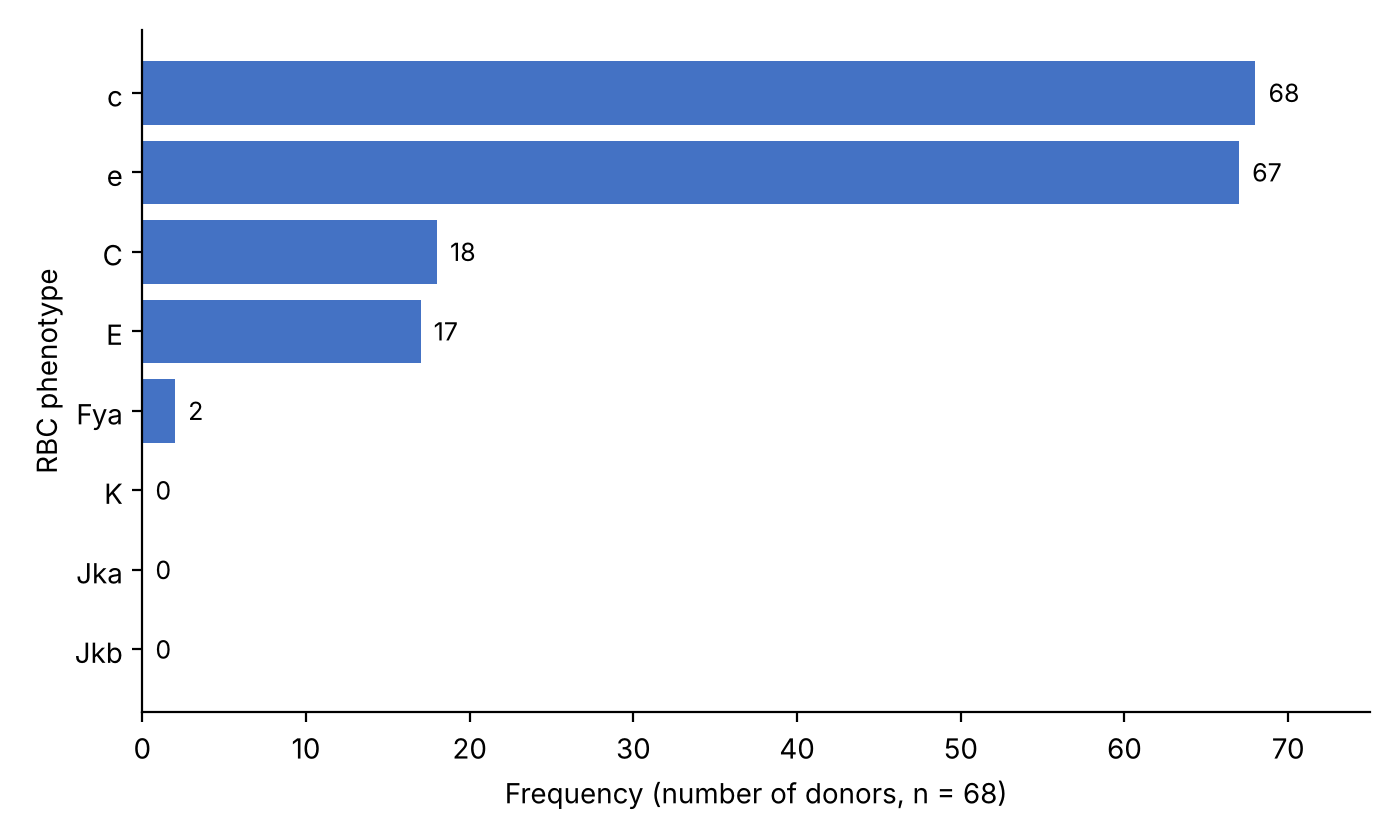
A total of 68 donors were recruited for the study. The mean age of the study participants was 30.6 ± 9.9. Majority of them were males 60 (88.2%) and 44 (64.7%) were less than 30 years old. The most prevalent red cell antigen was c (100%), followed by e antigen 67 (98.5). Only one of the blood donors lacked the e antigen. About a quarter had C 18 (26.5) and E 17 (25.0), while only 2(2.9%) donors had Fya. None of the donors had KEL1, JKa, and JKb antigens. (Tables 1 and 2). Figure 1 shows a pictorial representation of the distribution of red cell antigens in the study population.
Amongst the quarter of the study participants that possessed the C antigen, 15 (25.0) of them were males, and there was no statistically significant association between the red cell phenotypes and gender for the C antigen (p=0.428). None of the female donors had the Fya antigen when compared to the males, but this was not statistically significant. (p=1.000). There was an equal distribution across both genders for the other red cell antigens, but these were not statistically significant as seen in table 3. (p=1.000, 1.000) respectively.
| Age group | n (%) |
|---|---|
| <30 | 44 (64.7) |
| 31 – 40 | 12 (17.6) |
| 41 – 50 | 9 (13.2) |
| 51 – 60 | 3 (4.4) |
| Mean ± SD | 30.6 ± 9.9 |
| Range | 20 – 60 |
| Sex | |
| Male | 60 (88.2) |
| Female | 8 (11.8) |
| RBC extended phenotype | Frequency (n) Percentage (%) | 95% CI (%) |
|---|---|---|
| KEL1 | 0(0.0%) | 0.0–5.3 |
| C | 18(26.5%) | 16.4-38.6 |
| c | 68(100%) | 94.7-100 |
| E | 17(25%) | 15.3-36.9 |
| e | 67(98.5%) | 92.1-100.0 |
| JKa | 0(0.0%) | 0.0-5.3 |
| JKb | 0(0.0%) | 0.0-5.3 |
| FYa | 2(2.9%) | 0.4-10.2 |
| RBC extended phenotype distribution | Male n = 60 |
Female n = 8 |
p value |
|---|---|---|---|
| KEL1 | 0 (0.0%) | 0 (0.0%) | |
| C | 15 (25.0%) | 3 (37.5%) | 0.428 |
| c | 60 (100.0%) | 8 (100.0%) | NA |
| E | 15 (25.0%) | 2 (25.0%) | 1.000 |
| e | 59 (98.3%) | 8 (100.0%) | 1.000 |
| Jka | 0 (0.0%) | 0 (0.0%) | |
| Jkb | 0 (0.0%) | 0 (0.0%) | |
| Fya | 2 (3.3%) | 0 (0.0%) | 1.000 |
Note: Fisher’s exact test. NA = not applicable.
4Discussion
Blood transfusion remains a vital aspect of patient care. Devising a means of reducing red cell alloimmunization amongst multiply transfused patients is very crucial so that the patients may benefit maximally from transfused blood.
The Rh system is highly immunogenic and comprises of 5 major antigens: D,C, E, c, e. Rh positive or Rh negative is used to report the presence or absence of the D antigen. Clinically significant red cell antibodies are antibodies that are associated with hemolytic disease of the newborn, hemolytic transfusion reaction, which result in reduced red cell survival. Anti D is the most common cause of hemolytic disease of the newborn followed by anti c, which is the clinically most important Rh antigen after D [6]. It is therefore very essential to pay attention to the medical history of patients as it pertains to blood transfusion, pregnancies etc, before doing antibody identification test.
The findings of this study show a varied distribution of clinically significant red cell antigens in blood donors within a setting in Nigeria. Among 68 donors that were recruited in the research, the most prevalent red cell antigen was c, which was seen in all those tested, followed by e (98.5%). This study is similar to the study performed by Adewoyin et al. [8] in the south western part of Nigeria who reported that the prevalence of c and e antigens were 97.7% and 97.4%of their study population respectively. Considering the high immunogenicity of c, the high prevalence of c becomes highly beneficial as it markedly reduces the risk of alloimmunization in the general public. About a quarter of the study population were positive of C and E antigens. This poses a high risk of alloimmunization with these antigens because while up to a quarter of the population had the antigen, a larger population lack the antigen putting them at risk of alloimmunization. This corroborates with the studies done on the prevalent alloantibodies amongst multiply transfused patients, it was found that the most common alloantibodies were anti E, and anti C, followed by others [9-11]. Foreign studies showed varying prevalence in alloantibody formation with varying red cell antigens [12-15]. This necessitates production of antigram for antibody identification tailored to the antigenic peculiarities of a given population.
Adewoyin et al. [8] also observed a similar red cell antigen distribution as seen in our study. A related study performed amongst Ibibios, Efik and Igbos, showed similar findings with ours, suggesting some likely genetic similarities amongst the study group [16]. We also had similar findings with a study done in Côte d’Ivoire [17]. While our study recorded comparable findings in the prevalence of e and c antigens with some authors in Riyadh, they had a markedly higher prevalence of C antigen of up to 66% [18]. These differences further buttress the need to establish a population-based database.
We reported that none of the study participants had the KEL1 antigen. According to a study conducted amongst the Igbo tribe in Port Harcourt Nigeria, it reported that the KEL1 antigen is rare amongst the Igbo tribe [19]. A related study also showed that KEL1 antigen is rare in the Ikwerre indigenes of Rivers state [20]. Additionally, a study conducted among women of Lelna ethnic in northern Nigeria reported KEL1 antigen to be 6.6% among the study participants [21]. Christian et al. reported the absence of KEL1 antigen amongst Ijaw descendants living in Bayelsa state, Nigeria [22], while a study in south western Nigeria and northern Nigeria reported the prevalence of KEL 1 antigen as 6.0% and 2.0% respectively [23,24]. It is important to note that in this study conducted in Northern Nigeria, that the prevalence of KEL1 antigen was significantly higher among the Hausa ethnic group compared to the other ethnic groups. From literature, it is obvious that there may be certain genetic differences amongst the different tribes in Nigeria which may explain why there is rarity of KEL1 antigen in Igbos and a higher prevalence in non Igbos. This maybe due to evolutionary genetic variations. It is also good to note that our study was carried out amongst the Igbos in the southeastern part of Nigeria and that k antigen (KEL2 or cellano) was not tested for in our study. This would have given a more accurate prevalence of the K antigen. Onyenekwe et al. [25] in their study in Asaba and Awka in the southern southern and southeastern part of Nigeria also report only 2.1% prevalence of KEL1 antigen in their study group, which further suggests a low prevalence of KEL1 antigen in southern eastern Nigeria.
The Kidd glycoprotein is a multipass protein that functions as a urea transporter. Antibodies against the Kidd antigens are a notable cause of delayed hemolytic transfusion reactions. Anti-Kidd antibodies have also been implicated in the hemolytic disease of the newborn (HDN). The severity of the disease varies but tends to be mild in nature [26].
We found that none of the blood donors tested had Kidd antigens both Jka and JKb, which was unusual considering available literature. Several local studies reported varying findings. The distribution of Kidd antigens among pregnant women in sokoto studied indicated a prevalence of Jka, Jkb and Jk(a+b+) of 8 (4.9%), 13 (8.0%) and 0 (0.0%), respectively [27]. A study conducted in Rivers state Nigeria showed the distribution of kidd blood group among diabetic groups to be Jka+>Jkb+> Jkb- >Jka-is (100%, 94.7%, 5.3%, 0.00 ) respectively [28]. Some authors that studied the prevalence of kidd (JKa) antigens on the indigenes of Bonny Kingdom in Nigeria reported a low prevalence rate of 3.32% [29]. Similarly, a study done in Malawi showed that 98.2% of their study population were negative for the kidd antigen [30]. Our findings seem unusual as none of our study participants had neither Jka or Jkb as seen in other studies. This may probably be due to the failure of the kidd antisera. It is important to note that the test was carried out according to the manufacturer’s instruction.
Contrary to our findings, studies done in India, Maldives, Saudi Arabia and Korea showed a variable prevalence of the kidd antigen [31,32,33,34]. This suggests likely genetic differences across different races.
The Duffy blood group system is a complex of highly immunogenic glycoprotein antigens found on the surface of red blood cells (RBC), vasculature endothelial cells, alveolar epithelial cells, collecting tubules of the kidney, and on the surface of Purkinje cells in the brain. Antibodies against the Duffy blood group antigens are implicated in transfusion reactions and hemolytic disease of the fetus and newborn (HDFN) [36]. Aside being a blood group antigen, Duffy blood also as a receptor for a family of proinflammatory cytokines called chemokines, and is also a receptor for Plasmodium vivax malaria parasites. Therefore, individuals with the Duffy-negative phenotype are resistant to P. vivax invasion. Although P. vivax is widespread throughout the tropical and subtropical world, it is absent from West Africa, where more than 95% of the population is Duffy negative [36].
We reported a low prevalence of Fya (2.9%) in our study It is also important to note that we did not test for FyB because of lack of the antisera and financial constraints. This makes it difficult to make any inferences about the prevalence of duffy antigen amongst our study participants. A related study among pregnant women in Gombe state reported Fya of 6.6% [37]. The prevalence of Fya appears to be low in Nigeria, and this is worsened by the fact that there are a few previous studies to be referenced.
There was no statistically significant association in this study between the red cell phenotypes and gender for all the antigens tested, although a study reported female gender as a risk factor for development of red cell alloantibodies which is obviously because of pregnancies and subsequent transfusion risk and not necessarily a direct gender related genetic differences [38].
5Conclusion
This study shows the distribution of clinically significant red cell antigens in blood donors in Nnewi, Anambra State, Nigeria. Kell(K1) antigen was not present on any tested donors and there was no associated gender-based relationship between the red cell phenotypes distribution. Considering the racial peculiarities in red cell antigen expression, it is important to design the antigram based on the prevalent antigens in a given place. There is need for a more broad-based and extensive red cell phenotyping to help boost the information on red blood cell antigen expression obtained from this study. This will help in developing a local reagent red cell panel for antibody screening and identification, especially for the multiply transfused patients. This will help mitigate red cell alloimmunization and hemolytic blood transfusion reactions by ensuring these patients are given appropriately matched blood.
The study was limited by its small sample size and inadequate antisera, which made it difficult for us to achieve our minimum sample size. We fell short by 3 donors. We therefore recommend a more broad-based study to validate the findings of our study.
Funding
The research did not receive any form of funding but the antisera was a donation
Conflict of interest
The authors declare no conflict of interest
Data availability
Data will be made available on request
References
- [1]Al-Riyami AZ, Daar S. Transfusion in haemoglobinopathies: Review and recommendations for local blood banks and transfusion services in Oman. Sultan Qaboos Univ Med J. 2018;18(1):e3–e12. https://doi.org/10.18295/squmj.2018.18.01.002
- [2]Lotterman S, Sharma S. Blood transfusion [updated 2023 Jun 20]. In: StatPearls [Internet]. Treasure Island (FL): StatPearls Publishing; 2024. https://www.ncbi.nlm.nih.gov/books/NBK499824/
- [3]Erhabor O, Erhabor T, Adias TC, Polycarp II. Distribution of clinically relevant blood group antigens among Nigerians and the management of rhesus D negative pregnancies: Implications for haemolytic disease of the foetus and newborn and haemolytic transfusion reactions. IntechOpen; 2021. https://doi.org/10.5772/intechopen.90372
- [4]Enosolease ME, Bazuaye GN. Distribution of ABO and Rh-D blood groups in the Benin area of Niger-Delta: Implication for regional blood transfusion. Asian J Transfus Sci. 2008;2(1):3–5. https://doi.org/10.4103/0973-6247.39502
- [5]Lamba DS, Kaur R, Basu S. Clinically significant minor blood group antigens amongst North Indian donor population. Adv Hematol. 2013;2013:215454. https://doi.org/10.1155/2013/215454
- [6]Chou ST, Westhoff CM. The Rh and RhAG blood group systems. Immunohematology. 2010;26(4):177–185.
- [7]Getu J, Fekadu U, Melatwork T. Assessment of minor blood group system antigens and their phenotype among voluntary blood donors in Ethiopian Blood and Tissue Bank Service, Addis Ababa, Ethiopia. Ethiop J Health Sci. 2023;33(5):816–820.
- [8]Adewoyin A, Lee G, Adeyemo T, Awodu O. Rh and Kell blood group antigen prevalence in a multi-ethnic cohort in Nigeria: Implications for local transfusion service. Immunohematology. 2018;34:61–65. https://doi.org/10.21307/immunohematology-2018-011
- [9]Idongesit SA, Archibong UH, Edeheudim DE. Prevalence and specificities of immune red cell antibodies in adult patients with sickle cell anaemia and blood donors in Uyo, South-South Nigeria: A case-control study. Saudi J Pathol Microbiol. 2022;7(11):421–427.
- [10]Ayodeji OO, Nyong IC, Adams EE, Akinloye BO, Kosamat YA, Okolo CS. Prevalence of alloantibodies associated with haemolytic disease of the fetus and newborn in pregnant women at the Ekiti State University Teaching Hospital, Ado-Ekiti, Southwest Nigeria. Afr J Reprod Health. 2023;27(6s):70.
- [11]Ugwu NI, Awodu OA, Bazuaye GN, Okoye AE. Red cell alloimmunization in multi-transfused patients with sickle cell anemia in Benin City, Nigeria. Niger J Clin Pract. 2015;18(4):522–526.
- [12]Evers D, Middelburg RA, de Haas M, Zalpuri S, de Vooght KMK, van de Kerkhof D, et al. Red-blood-cell alloimmunisation in relation to antigens’ exposure and their immunogenicity: A cohort study. Lancet Haematol. 2016;3(6):e284–e292.
- [13]Amornrat VR, Piyapong S, Chalawan B, Ratchadaporn P, Ninnate J, Supawadee S. The prevalence, alloimmunization risk factors, antigenic exposure, and evaluation of antigen-matched red blood cells for thalassemia transfusions: A 10-year experience at a tertiary care hospital. Transfusion. 2019;59(1):177–184.
- [14]Zwingerman R, Jain V, Hannon J, Zwingerman N, Clarke G. Alloimmune red blood cell antibodies: Prevalence and pathogenicity in a Canadian prenatal population. J Obstet Gynaecol Can. 2015;37(9):784–790.
- [15]Thedsawad A, Taka O, Wanachiwanawin W. Prevalence and clinical significances of red cell alloimmunization and red cell bound immunoglobulin G in polytransfused patients with thalassemias. Hematology. 2019;24(1):208–214. https://doi.org/10.1080/16078454.2018.1549818
- [16]Jeremiah ZA, Odumody C. Rh antigens and phenotype frequencies of the Ibibio, Efik, and Ibo ethnic nationalities in Calabar, Nigeria. Immunohematology. 2005;21:21–24.
- [17]Bogui S, Dembele B, Sekongo Y, Abisse S, Konaté S, Sombo M. Phenotypic profile of Rh and Kell blood group systems among blood donors in Côte d’Ivoire, West Africa. J Blood Transfus. 2014;2014:309817.
- [18]Alalshaikh M, Almalki Y, Hasanato R, Almomen A, Alsughayir A, Alabdullateef A, et al. Frequency of Rh and K antigens in blood donors in Riyadh. Hematol Transfus Cell Ther. 2022;44(4):555–559. https://doi.org/10.1016/j.htct.2021.03.003
- [19]Serekara GC, Echonwere-Uwikor BE, Opara FA. Kell blood group antigen: Rare amongst Igbo descents in Nigeria. Arch Hematol Blood Dis. 2025;7(1):25–28.
- [20]Serekara GC, Echonwere-Uwikor BE, Celestine JS. Frequency of Kell (K) blood group antigen amongst indigenes of Ikwerre ethnicity in Rivers State, Nigeria. SJMLS. 2025;10(1).
- [21]Imoru M, Babangida S, Erhabor O, Mohammed H, Egeonu S. Distribution of ABO, rhesus phenotypes, and Kell blood group antigens among pregnant women of Lelna ethnic group in Kebbi State, Northwest Nigeria. J Hematol Allied Sci. 2025;5:66–71.
- [22]Christian SG, Igbogi A. Prevalence of Kell (K) antigen amongst Ijaw descents in Bayelsa State. BJMLS. 2025;10(2):1–8.
- [23]Nurudeen YO, Igbeneghu C, Oyeleke KO, Nassir SA. Assessing ABO and Kell blood group phenotypes and antigens in donated blood units at University College Hospital, Ibadan, Nigeria. Int Blood Res Rev. 2024;15(4):16–24.
- [24]Buhari H, et al. Blood group antigens phenotype frequencies among pregnant women in Sokoto State, Nigeria. Int Blood Res Rev. 2022;13(4):134–140.
- [25]Onyenekwe CC, et al. Occurrence of Kell blood group among individuals with ABO and Rh (D) blood groups in Awka and Asaba, Nigeria. Int J Biol Chem Sci. 2023;17(5):1797–1803.
- [26]Lawicki S, Covin RB, Powers AA. The Kidd (JK) blood group system. Transfus Med Rev. 2017;31(3):165–172.
- [27]Erhabor O, Hassan M, Alhaji YB, Yakubu A, Buhari H. Kidd blood group phenotypes among pregnant women in Sokoto, North Western Nigeria. Asian Pac J Trop Med. 2014;7S1:S111–S115. https://doi.org/10.1016/S1995-7645(14)60215-7
- [28]Joseph E, Serekara GC, Baribefe K. The prevalence of Kidd, Lewis, ABO and RhD blood group systems in diabetic patients attending RSUTH. Annals of Innovation in Medicine. 2025;3(1).
- [29]Jacob RB, Akah MN, Robinson-Mbato L, Christian SG, Olusola OA, Eze EM. Expression of Kidd, Rh-C, E, and D antigen among indigenes of Bonny Kingdom, Nigeria. J Blood Disord Transfus. 2022;13:492.
- [30]M’baya B, Mfune T, Mogombo E, Mphalalo A, Ndhlovu D, Knight RC. The prevalence of red cell antigens and antibodies in Malawi. Transfus Med. 2010;20(3):196–199.
- [31]Makroo RN, Bhatia A, Gupta R, Phillip J. Prevalence of Rh, Duffy, Kell, Kidd & MNSs blood group antigens in the Indian blood donor population. Indian J Med Res. 2013;137(3):521–526.
- [32]Saleem M, Ibrahim M. Characterization of Rh and other blood group systems amongst the Maldivian blood donors. Med J Malaysia. 2013;68(5):393–396.
- [33]Kuriri FA, Ahmed A, Alhumud F, Alanazi F, Abdalhabib EK. Frequency of Duffy, Kidd, Lewis, and Rh blood group antigens and phenotypes among donors in the Al-Ahsa region, Saudi Arabia. Clin Lab. 2024;70(7):1316.
- [34]Bae CO, Kwon SS, Kim S. Investigation of blood group genotype prevalence in Korean population using large genomic databases. Sci Rep. 2023;13:15326.
- [35]Neote K, Mak JY, Kolakowski LF, Schall TJ. Functional and biochemical analysis of the cloned Duffy antigen: Identity with the red blood cell chemokine receptor. Blood. 1994;84(1):44–52.
- [36]Miller LH, Mason SJ, Clyde DF, McGinniss MH. The resistance factor to Plasmodium vivax in blacks. The Duffy-blood-group genotype, FyFy. N Engl J Med. 1976;295(6):302–304.
- [37]Gaji AM, Ogbenna A, Girei AI, Pindiga KM, Adamu S, Yuguda S, et al. Duffy red cell antigen phenotype among indigenous pregnant women attending antenatal clinic at Federal Teaching Hospital Gombe, Nigeria. Afr J Lab Haem Transf Sci. 2023;2(4):266–276.
- [38]Verduin EP, Brand A, Schonewille H. Is female sex a risk factor for red blood cell alloimmunization after transfusion? A systematic review. Transfus Med Rev. 2012;26(4):342–353.e5.