Pharmacy
Bioassay-Guided Isolation and Characterization of Anti-Inflammatory Constituents from Mimosa pudica L. Leaves
Department of Pharmacognosy, Institute of Pharmaceutical Science and Research, Unnao, Uttar Pradesh, India (affiliated to Dr. A. P. J. Abdul Kalam Technical University, Lucknow, Uttar Pradesh, India)
Published October 2026
Abstract
Chronic, non-resolving inflammation is now recognised as the shared pathophysiological substrate of most major non-communicable diseases, yet the long-term use of non-steroidal anti-inflammatory drugs (NSAIDs), glucocorticoids and biologic agents remains constrained by gastrointestinal, cardiovascular, renal, metabolic and economic liabilities. Mimosa pudica L. (Fabaceae), the "sensitive plant", carries an unusually consistent cross-cultural record of use for inflammatory and rheumatic complaints in the Ayurvedic, Siddha, Unani, Traditional Chinese and Latin American systems of medicine. The present study applied a complete bioassay-guided fractionation cycle to identify and pharmacologically characterise the principal anti-inflammatory constituent of M. pudica leaves. Authenticated, shade-dried leaf material (voucher MP-WG-2024–17) was standardised against Indian Pharmacopoeia specifications and subjected to sequential Soxhlet extraction with six solvents of ascending polarity (n-hexane, chloroform, ethyl acetate, ethanol, methanol and water), giving yields of 2.84–18.45 % w/w. Triage of the six fractions by DPPH and ABTS radical-scavenging and by hypotonic-solution-induced human red blood cell (HRBC) membrane stabilisation identified the ethyl acetate fraction as the most active (DPPH IC50 = 21.4 ± 1.3 µg/mL; HRBC stabilisation 71.6 ± 3.4 % at 200 µg/mL). Silica-gel column chromatography of this fraction with in vivo screening of the pooled sub-fractions, followed by Sephadex LH-20 gel filtration and preparative thin-layer chromatography, delivered 142 mg of a yellow crystalline solid designated MP-C1 (HPLC-DAD purity 98.7 %). Integrated UV-Visible, FT-IR, 1H and 13C NMR and high-resolution electrospray ionisation mass spectrometry unambiguously identified MP-C1 as quercetin (3,3′,4′,5,7-pentahydroxyflavone; C15H10O7; MW 302.24 g/mol). Acute oral toxicity testing under OECD Test Guideline 423 showed no mortality at the 2000 mg/kg limit dose (LD50 > 2000 mg/kg). In carrageenan-induced rat paw oedema, quercetin at 100 mg/kg p.o. produced 63.0 ± 1.6 % inhibition at the four-hour peak, statistically indistinguishable from diclofenac sodium at 10 mg/kg (67.8 ± 1.4 %; p > 0.05), with an ED50 of approximately 60 mg/kg. In the seven-day cotton-pellet granuloma model, quercetin at 100 mg/kg inhibited granuloma formation by 42.5 ± 1.9 %, exceeding diclofenac (35.4 ± 1.7 %). Quercetin lowered serum TNF-α, IL-6 and CRP by 46–50 % and restored tissue malondialdehyde, glutathione, superoxide dismutase and catalase substantially closer to normal than did diclofenac (mean residual redox deficit 11 % versus 23 %). Histopathology confirmed tissue protection with no hepatic or renal injury. The resulting therapeutic index above 30 identifies M. pudica leaf-derived quercetin as a multi-target, multi-mechanism anti-inflammatory bioactive warranting formulation-directed translational development.
Keywords: Mimosa pudica; quercetin; bioassay-guided fractionation; anti-inflammatory activity; carrageenan paw oedema; cotton-pellet granuloma; TNF-α; oxidative stress; NF-κB; phytopharmacology
1Introduction
1.1Chronic inflammation as a twenty-first-century therapeutic target
Inflammation is the most fundamental and evolutionarily conserved response of multicellular organisms to tissue insult. Classically defined by Celsus through the cardinal signs of rubor, tumor, calor and dolor, to which Virchow added functio laesa, it was for many centuries regarded as a discrete, self-limiting reaction whose purpose was the elimination of pathogens, the clearance of damaged cells and the restoration of tissue homeostasis [1]. The past three decades have produced a profound conceptual recalibration. Low-grade, chronic, non-resolving inflammation is now understood to be the unifying pathophysiological substrate of most non-communicable diseases, including atherosclerotic cardiovascular disease, type-2 diabetes mellitus, the metabolic syndrome, chronic obstructive pulmonary disease, osteoarthritis, rheumatoid arthritis, the autoimmune spectrum, several neurodegenerative conditions and a substantial fraction of solid and haematological malignancies [1–4].
The magnitude of the resulting burden is difficult to overstate. Cardiovascular disease alone accounts for approximately 17.9 million deaths annually, a majority traceable to chronic vascular inflammation as the proximal driver [3], and Global Burden of Disease analyses attribute more than half of all-cause adult mortality worldwide to processes in which non-resolving inflammation plays a critical or contributory role [4]. Type-2 diabetes mellitus, affecting more than 537 million people and projected to reach 783 million by 2045, is now characterised as a meta-inflammatory disorder driven by adipose-tissue cytokine dysregulation and persistent NF-κB activation [2,4], while inflammatory bowel disease, asthma, atopic dermatitis, psoriasis, lupus and multiple sclerosis together affect over 700 million individuals with rising incidence in both high- and low-income settings [4,5].
Mechanistically, what distinguishes chronic from acute inflammation is the failure of dedicated pro-resolving programmes that normally terminate the response within days through specialised mediators - lipoxins, resolvins, protectins and maresins - and through the M1-to-M2 macrophage polarisation switch [2]. When resolution fails, persistent activation of innate immune cells, sustained engagement of pattern-recognition receptors, constitutive NF-κB signalling and dysregulated cytokine networks (TNF-α, IL-1beta, IL-6, IL-17, IFN-γ) drive a self-perpetuating cycle of tissue damage, oxidative stress, fibrosis and organ dysfunction [1,2,5]. Because every step of this cascade is modulated by cellular redox state, reactive oxygen and nitrogen species act simultaneously as effectors of injury and as second messengers activating NF-κB, AP-1 and HIF-1alpha. Pharmacological intervention must therefore contend with selectivity, pathway redundancy and phase-dependence at once.
1.2Persistent limitations of conventional anti-inflammatory pharmacotherapy
Despite four decades of intensive investment, the clinical armamentarium remains dominated by a few drug classes, each constrained by safety, tolerability or cost. Long-term NSAID exposure carries an annual incidence of peptic ulcer disease of 1–4 % with overt gastrointestinal bleeding in 1–2 % of users [6,7]. The attempt to decouple efficacy from gastrotoxicity through COX-2-selective inhibition proved only partially successful: suppression of endothelial prostacyclin without proportionate reduction of platelet thromboxane A2 conferred increased thrombotic risk, culminating in the withdrawal of rofecoxib and regulatory recalibration of the entire coxib class [6–8]. Renal complications affect 1–5 % of long-term users [6].
Glucocorticoids remain the most powerful broad-spectrum agents available, acting through receptor-mediated transactivation of anti-inflammatory genes and transrepression of NF-κB and AP-1 [9]. Their potency is paralleled by an extensive spectrum of dose- and duration-dependent adverse effects that in long-term use are essentially universal: iatrogenic Cushing syndrome, glucose intolerance, osteoporosis, hypertension, opportunistic infection, cataract, neuropsychiatric disturbance, impaired wound healing and adrenal suppression [9]. Biological and targeted synthetic disease-modifying agents have transformed outcomes in severe disease but are limited by parenteral administration, immunogenicity, serious opportunistic infection and acquisition costs beyond reach for most patients in low- and middle-income settings [9]. Even in well-resourced systems, fewer than half of patients with chronic inflammatory disease achieve sustained remission, and adverse-event-driven discontinuation exceeding 30 % at five years is commonplace. This therapeutic gap motivates the renewed search for safer, multi-target, naturally occurring anti-inflammatory agents.
1.3Natural products and the ethnopharmacological rationale
The contribution of natural products to the modern drug supply remains extraordinary. In their landmark periodic analysis of the 1562 small-molecule new chemical entities approved between January 1981 and December 2014, Newman and Cragg concluded that approximately 51 % were unaltered natural products, semi-synthetic derivatives, synthetic mimics retaining a natural-product pharmacophore, or compounds inspired by a natural-product lead [10]. The dependence is more pronounced still in specific therapeutic areas: roughly 83 % of approved anti-cancer agents, 73 % of anti-infectives and 64 % of anti-inflammatory drugs are natural-product derived or inspired [10]. Several intrinsic features account for this disproportionate productivity - structural diversity exceeding that of synthetic combinatorial libraries, evolutionary pre-selection for interaction with biological macromolecules, molecular geometry complementary to protein binding sites, and a marked tendency toward poly-pharmacology that is increasingly recognised as advantageous in diseases of multifactorial origin [11–13].
The World Health Organization estimates that approximately 80 % of the global population relies on traditional medicine, predominantly plant-based, as a primary or exclusive modality of healthcare [14]. Ethnobotanical leads carry a documented advantage in early-stage discovery: prospective screening of plants used traditionally for a specific indication yields hit rates several-fold higher than random screening of unselected species [15]. India, with its codified Ayurvedic, Siddha and Unani traditions and some 7500 documented medicinal species, is among the world’s richest settings for such discovery.
1.4Mimosa pudica L.: botany, ethnomedicine and phytochemistry
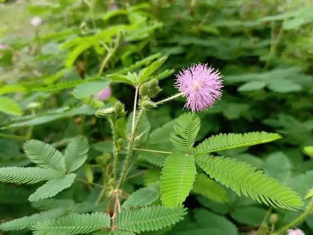
Mimosa pudica L. is a small, prostrate-to-ascending herbaceous perennial of the family Fabaceae, subfamily Mimosoideae, native to the tropical Americas and now pantropical in distribution [16,17]. The specific epithet pudica (Latin: shy, modest) reflects the plant’s most arresting characteristic, the rapid and reversible thigmonastic folding of its compound leaflets within one to two seconds of mechanical, thermal or electrical stimulation, mediated by an action-potential-like wave propagating along the rachis at 1–3 cm/s and by rapid efflux of K+, Ca2+ and Cl- from pulvinar motor cells [18]. In the Indian subcontinent the plant occurs as a common weed of disturbed roadsides, fallow fields and plantation understoreys from sea level to about 1300 m [19,20].
The species occupies an established position in the materia medica of every major tropical medical tradition. In Ayurveda, where it is known as Lajjalu, it is classified as kasaya (astringent) and tikta (bitter) with sheeta (cold) virya and is prescribed for bleeding disorders, dysentery, piles, fistula, vrana (ulcers and wounds), diabetes, gynaecological conditions, asthma, fever and rheumatic complaints [19,20]. Siddha and Unani practice employ it for piles, gonorrhoea, leprosy, skin disease and ulcers [21]. In Traditional Chinese Medicine, Han Xiu Cao is regarded as bitter, slightly cold and slightly toxic, entering the heart and liver meridians to clear heat, calm the spirit, reduce swelling and detoxify [22,23]. Latin American, sub-Saharan African and South-East Asian traditions converge on the same two dominant applications: anti-inflammatory and wound-healing use, and antimicrobial use against a broad spectrum of bacterial and fungal pathogens [20,24,34].
Half a century of phytochemical investigation has established M. pudica as a chemically rich species containing seven principal classes of secondary metabolite. Among flavonoids, quercetin (3,3′,4′,5,7-pentahydroxyflavone) is the principal aglycone, accompanied by kaempferol, luteolin, apigenin, rutin and isoquercitrin, with leaves and flowers carrying the highest concentrations [22,23,24]. Mimosine, the signature non-protein amino acid of the genus, accumulates in seeds and roots [25]. Phenolic acids (gallic, caffeic, chlorogenic, ferulic, p-coumaric, protocatechuic), condensed catechin- and gallocatechin-derived tannins dominating the root system, triterpenoid saponins of the oleanane and ursane series including mimonosides A-C, and the sterols β-sitosterol and stigmasterol complete the inventory [26–29,35]. These classes map onto the six recognised flavonoid subclasses and the wider polyphenol framework that governs their pharmacology [36,37]. The pharmacological spectrum reported for the species is correspondingly broad, encompassing anti-inflammatory, antioxidant, antimicrobial, wound-healing, hepatoprotective, antidiabetic, antinociceptive, anxiolytic, antivenom and selected anticancer activities [19,20,30,31].
1.5Rationale, hypotheses and objectives of the present study
Three converging lines of evidence make the anti-inflammatory pharmacology of M. pudica a compelling object of investigation. First, the burden of inflammation-driven disease and the limitations of conventional pharmacotherapy establish a clear unmet need. Second, the chemical composition of the species is dominated by precisely those polyphenolic constituents - flavonoids, phenolic acids and tannins - most extensively validated as anti-inflammatory and antioxidant pharmacophores [29]. Third, the ethnomedical record for inflammation-related conditions is exceptionally consistent across geographically and culturally independent traditions, satisfying the criterion of cross-cultural convergence that ethnopharmacologists regard as the strongest empirical predictor of pharmacological validity [19,20,21].
Nevertheless, several aspects of this pharmacology remain insufficiently consolidated. Earlier investigations have reported paw-oedema inhibition in the range of 50–65 % and granuloma reduction of 35–45 % using crude leaf or root preparations at 200–400 mg/kg [30–33], but the relative contribution of individual phytochemicals to the activity of crude extracts has been incompletely defined; mechanistic studies have been conducted in fragmentary fashion across many laboratories with limited integration; and the cytokine and redox correlates of the macroscopic anti-inflammatory response have rarely been documented within a single coherent experiment.
The present investigation was designed on two falsifiable hypotheses. First, that M. pudica leaves contain one or more flavonoid bioactives isolable as a single chemically pure entity by polarity-guided sequential extraction followed by bioassay-directed chromatography. Second, that the isolated bioactive would exhibit dose-dependent inhibition of both acute and chronic inflammation in standard rodent models, with magnitude comparable to a reference NSAID, achieved through measurable suppression of pro-inflammatory cytokines and restoration of endogenous antioxidant defences, within a safety margin substantially exceeding that of conventional anti-inflammatory drugs. Accordingly, the specific objectives were to authenticate and pharmacognostically standardise the plant material; to perform sequential extraction and qualitative phytochemical screening of all fractions; to triage the fractions by in vitro antioxidant and membrane-stabilisation assays; to isolate and structurally characterise the principal bioactive; to determine acute oral toxicity under OECD Test Guideline 423; and to evaluate acute and chronic anti-inflammatory efficacy against diclofenac sodium, supported by serum cytokine profiling, tissue oxidative-stress measurement and histopathology.
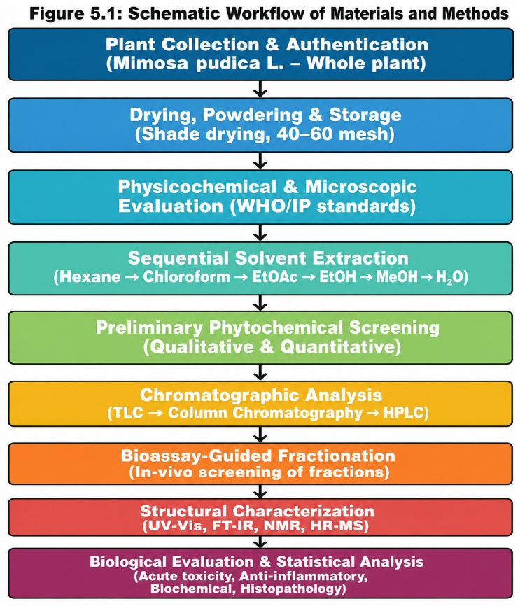
2Materials and methods
2.1Chemicals, reagents and instrumentation
All extraction and chromatographic solvents were of analytical reagent grade and redistilled before use; HPLC-grade solvents were from Merck (Darmstadt, Germany). Silica gel (60–120 and 230–400 mesh), pre-coated silica gel 60 F254 TLC plates (Merck), Sephadex LH-20, Folin-Ciocalteu reagent, DPPH, ABTS, quercetin reference standard (≥ 95 %, Sigma-Aldrich) and Dragendorff reagent were from authorised suppliers; lambda-carrageenan (Type IV, Sigma-Aldrich) served as phlogogen and diclofenac sodium as reference drug. Instrumentation comprised a Borosil Soxhlet assembly, Buchi R-300 rotary evaporator, Shimadzu UV-1800 spectrophotometer, Shimadzu IR-Affinity-1S FT-IR spectrophotometer, Shimadzu LC-2030C Plus HPLC with photodiode-array detector, Bruker Avance III 400 MHz NMR spectrometer, Waters Xevo G2-XS QTof mass spectrometer, Ugo Basile Model 7140 plethysmometer, Leica RM 2125 RTS microtome and Bio-Rad iMark plate reader. Measurements were made at 24 ± 2 °C and 45–55 % relative humidity.
2.2Plant material, authentication and processing
Mimosa pudica L. material was collected at the peak vegetative to early flowering stage (September-October 2024) from authenticated wild populations at 31 degrees 06′ N, 77 degrees 10′ E and approximately 1150 m above mean sea level, on well-drained sandy-loam soil of pH 6.2–6.8. Sites were selected for absence of agrochemical contamination and collection conducted under the WHO guidelines on Good Agricultural and Collection Practices [45]. Approximately 8.0 kg of healthy material was harvested with sterilised tools and transported under cold chain within four hours. Botanical authentication was performed by a qualified taxonomist using standard keys and the diagnostic characters of the species - bipinnately compound seismonastic leaves, pink globose capitate inflorescences, recurved stem prickles and transversely segmented prickly legumes - in accordance with WHO guidelines for research on traditional medicine [15], and a voucher specimen (MP-WG-2024–17) was deposited in the institutional herbarium.
Material was washed, rinsed with distilled water and shade-dried at 24 ± 2 °C under controlled ventilation for 14–18 days; shade drying was preferred to oven or sun drying to prevent thermal degradation of thermolabile constituents and photochemical decomposition of flavonoids [46]. The dried material was ground and sieved through a No. 40 sieve to a uniform 40–60 mesh particle size, critical for reproducible extraction efficiency, and stored in airtight amber glass protected from light. The total yield of dry powder was 1.42 kg, approximately 17.7 % of fresh weight.
2.3Pharmacognostic and physicochemical evaluation
Organoleptic characters of fresh and dried material were documented. Free-hand transverse sections (8–12 µm) of root, stem and leaf were cleared in chloral hydrate, stained with safranin and fast green and examined at 100x, 400x and 1000x, and powder microscopy performed on the drug in glycerol. Loss on drying, total ash, acid-insoluble ash, water-soluble ash, water- and alcohol-soluble extractive values, foreign organic matter and pH of a 1 % w/v aqueous suspension were determined in triplicate per the Indian [47], British [48] and European [49] Pharmacopoeias.
2.4Sequential solvent extraction
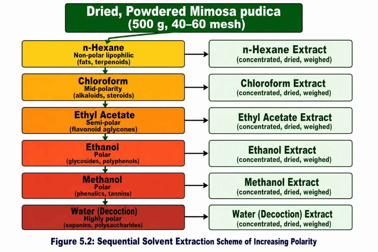
A polarity-graded sequential strategy was adopted to permit selective recovery of compound classes from lipophilic terpenoids to polar saponins, and to defat the drug early so that polar bioactives concentrate in later fractions [50,51]. Six solvents were used in ascending polarity (Figure 3). Hot continuous Soxhlet extraction was applied to n-hexane, chloroform and ethyl acetate, using 50 g of powder with 500 mL of solvent for 8 hours or until the siphon draining became colourless. Cold maceration was used for ethanol and methanol, both efficient vehicles for thermolabile flavonoid glycosides that Soxhlet conditions may degrade: 100 g of powder was macerated with 1000 mL of solvent for 72 hours with intermittent shaking and the marc re-extracted twice. The aqueous extract was prepared by decoction under reflux for 2 hours followed by lyophilisation. All extracts were filtered, concentrated under reduced pressure at 40 °C, dried to constant weight and stored at 4 degrees.
2.5Phytochemical screening
All six extracts, reconstituted at 10 mg/mL, were subjected to standard qualitative tests for alkaloids (Mayer, Wagner, Dragendorff), flavonoids (Shinoda, alkaline reagent), tannins and phenolics (ferric chloride, gelatin), saponins (foam), terpenoids (Salkowski), steroids (Liebermann-Burchard), glycosides (Keller-Killiani), carbohydrates (Molisch) and proteins (Biuret) following compendial procedures [46], graded on intensity of the diagnostic colour or precipitate. Total phenolic content was determined by the Folin-Ciocalteu method at 760 nm against gallic acid, total flavonoid content by the aluminium chloride method at 415 nm against quercetin, and total alkaloid content gravimetrically, all in triplicate.
2.6In vitro bioassay triage
Two mechanistically distinct endpoints were used to triage the six fractions and reduce assay-specific false positives, following the two-assay screening logic established for ethnopharmacological plant panels [57]. DPPH scavenging was measured after 30 minutes of dark incubation at 517 nm against ascorbic acid and BHT, and ABTS decolourisation at 734 nm, with IC50 values from non-linear regression. Membrane stabilisation was assessed by hypotonic-solution-induced human red blood cell haemolysis, with extract at 200 µg/mL and diclofenac sodium as reference. Assays were performed in triplicate.
2.7Bioassay-guided fractionation and isolation
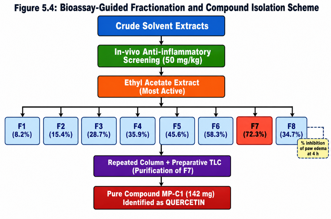
The ethyl acetate extract, identified as most active in the in vitro triage, was fractionated on a glass column (60 × 3 cm) wet-packed with 250 g of silica gel (60–120 mesh). Twenty-five grams of extract was adsorbed onto an equal weight of silica and eluted with a step-wise gradient of n-hexane, n-hexane:ethyl acetate (90:10 to 30:70), ethyl acetate, ethyl acetate:methanol (95:5 to 80:20) and methanol. The 156 fractions of 25 mL collected were monitored by TLC and pooled by profile into eight sub-fractions, F1–F8, each screened for anti-inflammatory activity in the carrageenan model at 50 mg/kg so that every fractionation step was anchored to biological evidence rather than chemical convenience [50,51]. The most active sub-fraction, F7, was purified by repeated chromatography on finer silica (230–400 mesh), Sephadex LH-20 gel filtration in methanol and preparative TLC, yielding 142 mg of a crystalline compound designated MP-C1, recrystallised twice from hot methanol (Figure 4).
Purity was established by analytical HPLC on a reversed-phase C18 column (250 × 4.6 mm, 5 µm, Phenomenex Luna) at 30 °C with 0.1 % v/v aqueous formic acid (A) and acetonitrile (B) at 1.0 mL/min, using a gradient from 10 % to 90 % B over 30 minutes with re-equilibration to 40 minutes, 20 µL injection of a 1 mg/mL solution and detection at 254 and 360 nm. The MP-C1 peak was identified by retention time, on-line UV spectrum and co-injection against an authentic quercetin standard, with the method validated under ICH Q2(R1) [63].
2.8Structural characterization
UV-Visible spectra were recorded over 200–600 nm in methanol (10 µg/mL), with bathochromic shift experiments using sodium methoxide, aluminium chloride, aluminium chloride/hydrochloric acid, sodium acetate and sodium acetate/boric acid to establish the hydroxylation pattern of the flavonoid skeleton. FT-IR spectra were acquired over 4000–400 cm−1 from a KBr pellet with 32 scans at 4 cm−1 resolution. One- and two-dimensional NMR spectra (1H, 13C, DEPT-135, COSY, HSQC, HMBC) were recorded at 25 °C in DMSO-d6 with tetramethylsilane as internal reference. High-resolution mass spectra were acquired in positive and negative electrospray modes with accurate mass within ± 5 ppm and tandem MS at 20 eV collision energy.
2.9Animals, ethics and acute oral toxicity
Adult male albino Wistar rats (180–220 g, 8–10 weeks) and Swiss albino mice were acclimatised for at least seven days at 24 ± 2 °C, 55 ± 10 % relative humidity and a 12-hour light/dark cycle with standard diet and water ad libitum except during brief overnight fasting. All procedures were approved by the Institutional Animal Ethics Committee and conducted under CPCSEA (Government of India) guidelines and the 3Rs principle, with design and reporting conforming to ARRIVE recommendations.
Acute oral toxicity of MP-C1 was determined under OECD Test Guideline 423, the acute toxic class method [58]. Animals (three per dose step) were fasted overnight and dosed by gavage with compound suspended in 0.5 % w/v sodium carboxymethylcellulose at successive starting doses of 5, 50, 300 and 2000 mg/kg. They were observed continuously for four hours for behavioural, autonomic and neurological signs and daily thereafter for 14 days, with body weight, intake and gross pathology recorded, and LD50 estimated from the OECD class-method decision tree.
2.10Carrageenan-induced paw oedema
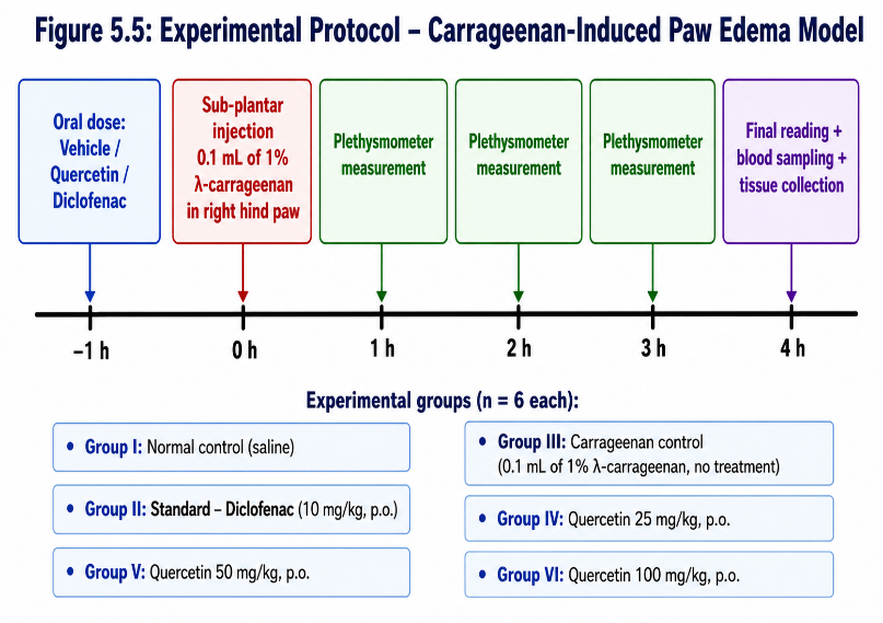
Acute anti-inflammatory activity was evaluated by the carrageenan-induced rat hind-paw oedema method of Winter and co-workers [52] following the standardised protocol of Morris [53]. The model is widely accepted because its cascade - histamine, serotonin and bradykinin early, prostaglandins, kinins and complement late - resembles human acute inflammation [55]. Animals were randomised into six groups of six: normal control (0.9 % saline p.o.), carrageenan control, diclofenac sodium 10 mg/kg p.o., and quercetin at 25, 50 and 100 mg/kg p.o. After overnight fasting, treatments were given orally in 0.5 % w/v carboxymethylcellulose exactly one hour before subplantar injection of 0.1 mL of 1 % w/v lambda-carrageenan into the right hind paw, the left paw serving as contralateral control [56]. Paw volume was measured by digital plethysmometry before injection and at 1, 2, 3, 4, 5 and 6 hours (Figure 5), and percentage inhibition calculated as [(Vc - Vt)/Vc] x 100.
2.11Cotton-pellet granuloma
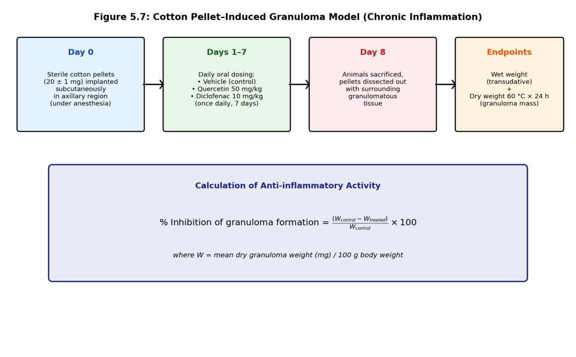
The proliferative phase of chronic inflammation was assessed by the cotton-pellet granuloma model of Swingle and Shideman [54]. Sterilised pellets (20 ± 1 mg, autoclaved at 121 °C for 15 minutes) were implanted subcutaneously, one in each axillary region, under diethyl ether anaesthesia. Animals were allocated to the same six groups and treated orally once daily for seven days from implantation. On day 8 the pellets with surrounding granulomatous tissue were dissected out, weighed wet as an index of transudate, dried at 60 °C for 24 hours and reweighed as an index of granuloma mass, with percentage inhibition calculated against inflammation-control values (Figure 6).
2.12Biochemical estimations and histopathology
Blood was collected from the retro-orbital plexus under light ether anaesthesia and centrifuged at 3000 × g for 10 minutes at 4 °C, and the serum stored at -80 °C. Serum CRP, TNF-α and IL-6 were quantified with rat-specific sandwich ELISA kits (Krishgen Biosystems, India) read at 450 nm. Paw tissue homogenates (10 % w/v in ice-cold 50 mM phosphate buffer, pH 7.4) were centrifuged at 10 000 × g for 15 minutes at 4 °C and assayed for malondialdehyde by the thiobarbituric-acid-reactive-substances method, reduced glutathione by the Ellman DTNB method, superoxide dismutase by pyrogallol auto-oxidation and catalase by the rate of decrease in hydrogen peroxide absorbance at 240 nm.
Paw, liver and kidney samples were fixed in 10 % v/v neutral buffered formalin, dehydrated through graded ethanol, cleared in xylene, embedded in paraffin and sectioned at 5 µm. Sections were stained with haematoxylin and eosin, with granulomatous tissue additionally stained by Masson trichrome for collagen, and evaluated at 100x and 400x for cellular infiltration, oedema, vascular congestion and organ-specific toxicity by a pathologist blinded to treatment allocation.
2.13Statistical analysis
Data are expressed as mean ± SEM for n = 6 animals per group, or mean ± SD for in vitro determinations in triplicate, and were analysed in GraphPad Prism 8.0 [60] and IBM SPSS Statistics 26.0 [61]. Normality and homogeneity of variance were verified by the Shapiro-Wilk and Levene tests. Multiple-group comparisons used one-way ANOVA with Dunnett or Tukey HSD post-hoc tests; time-course data were analysed by repeated-measures two-way ANOVA and dose-response curves fitted by four-parameter logistic regression. Differences with p < 0.05 were considered significant [59]. Analytical methods met ICH Q2(R1) criteria of linearity R2 ≥ 0.999, recovery 98–102 % and RSD < 2 % [63], and the quality framework conforms to FDA guidance for botanical drug products [62].
3Results
3.1Authentication and pharmacognostic standardisation
All collected specimens were authenticated as Mimosa pudica L. (Fabaceae), and the deposited voucher now serves as the permanent botanical reference for the study. Powder microscopy revealed paracytic stomata, multicellular uniseriate covering trichomes, abundant prismatic and cluster calcium-oxalate crystals, simple starch grains and lignified xylem vessels, all consistent with published descriptions and collectively sufficient to exclude adulteration with morphologically similar leguminous taxa. Quantitative pharmacognostic indices (Table 1) fell within Indian Pharmacopoeia acceptance ranges, indicating raw material that was clean, of low ash burden and rich in alcohol- and water-soluble constituents - qualities favourable for recovery of polyphenolic bioactives.
| Parameter | Observed value | Pharmacopoeial acceptance range |
|---|---|---|
| Loss on drying (% w/w) | 8.6 ± 0.3 | ≤ 10.0 |
| Total ash (% w/w) | 9.4 ± 0.4 | ≤ 12.0 |
| Acid-insoluble ash (% w/w) | 1.2 ± 0.1 | ≤ 2.5 |
| Water-soluble extractive (% w/w) | 18.6 ± 0.7 | ≥ 12.0 |
| Alcohol-soluble extractive (% w/w) | 16.4 ± 0.6 | ≥ 10.0 |
| Foreign organic matter (% w/w) | < 0.5 | ≤ 2.0 |
All determinations performed in triplicate according to Indian Pharmacopoeia monograph procedures [47].
3.2Sequential extraction yields and phytochemical profile
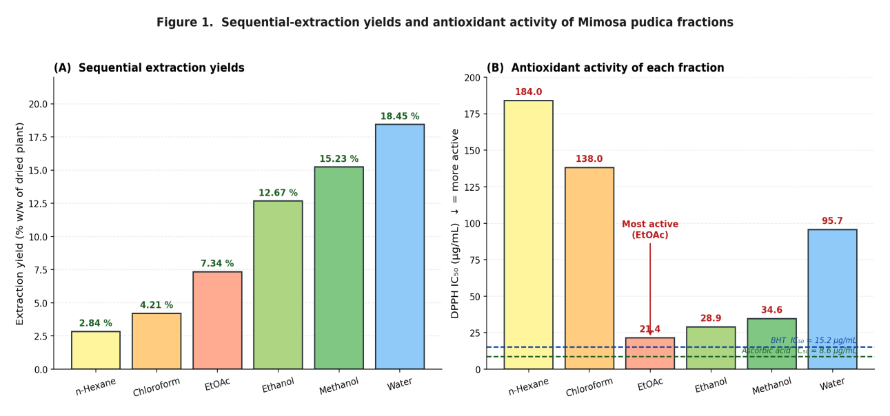
Sequential partitioning of 250 g of dried powder with six solvents of progressively increasing polarity gave the yield profile shown in Figure 7A. Recovery climbed monotonically from 2.84 % w/w in the most lipophilic n-hexane extract, through 4.21 % for chloroform, 7.34 % for ethyl acetate, 12.67 % for ethanol and 15.23 % for methanol, to 18.45 % w/w in the strongly polar aqueous fraction. This right-skewed distribution is characteristic of leguminous medicinal herbs whose chemical inventory is dominated by mid- to high-polarity phenolics, glycosides and water-soluble carbohydrates, and is broadly consistent with yields reported in independent investigations of the species [32,33].
Qualitative phytochemical screening (Table 2) confirmed the polarity-dependent partitioning the sequential design was intended to achieve. Steroids and terpenoids dominated the n-hexane and chloroform fractions, alkaloids appeared most strongly in chloroform, aglycone flavonoids and free phenolic acids concentrated in ethyl acetate, tannins and polar glycosides peaked in ethanol and methanol, and carbohydrates and protein-related material accumulated in the aqueous extract. Crucially, the ethyl acetate fraction alone gave intense positive Shinoda and aluminium chloride reactions for aglycone flavonoids without major interference from tannins or sugars, marking it as the most promising starting material for purification of a single active species.
| Constituent class | Hexane | CHCl3 | EtOAc | EtOH | MeOH | Water |
|---|---|---|---|---|---|---|
| Alkaloids | - | ++ | + | + | + | - |
| Flavonoids | - | + | +++ | ++ | ++ | + |
| Tannins / phenolics | - | - | + | ++ | +++ | ++ |
| Saponins | - | - | - | ++ | ++ | + |
| Steroids / terpenoids | ++ | +++ | + | - | - | - |
| Glycosides | - | + | + | +++ | ++ | + |
| Carbohydrates | - | - | - | + | ++ | +++ |
| Proteins / amino acids | - | - | - | - | + | ++ |
| Extractive yield (% w/w) | 2.84 | 4.21 | 7.34 | 12.67 | 15.23 | 18.45 |
Key: - absent; + weakly positive; ++ moderately positive; +++ strongly positive. Yields calculated on 250 g of dried powder.
3.3In vitro antioxidant and membrane-stabilisation triage
DPPH radical-scavenging served as the primary in vitro filter for prioritising the six extracts. As shown in Figure 7B and Table 3, the ethyl acetate fraction was the clear front-runner with an IC50 of 21.4 ± 1.3 µg/mL, a value approaching that of ascorbic acid (8.6 ± 0.4 µg/mL) and comparing favourably with the synthetic antioxidant BHT (15.2 ± 0.7 µg/mL) when normalised for molar mass. The ethanolic and methanolic fractions trailed at 28.9 ± 1.7 and 34.6 ± 2.0 µg/mL respectively, while the non-polar hexane and chloroform extracts and the aqueous fraction performed an order of magnitude worse. ABTS decolourisation produced an identical rank ordering.
The HRBC assay generated a parallel ranking, the ethyl acetate fraction protecting 71.6 ± 3.4 % of erythrocyte membranes at 200 µg/mL against 63.2 ± 3.0 % for ethanol, 57.5 ± 2.8 % for methanol, 38.4 ± 2.6 % for chloroform and 24.7 ± 2.1 % for hexane, relative to 82.4 ± 2.7 % for diclofenac. Because the erythrocyte membrane is functionally analogous to the lysosomal membrane, stabilisation is an accepted surrogate for inhibition of lysosomal enzyme release during inflammation. Convergence of two mechanistically distinct endpoints - one cell-free and radical-based, the other cellular and membrane-based - onto the same fraction justified advancing only the ethyl acetate extract to chromatography.
| Fraction / standard | DPPH IC50 (µg/mL) | ABTS IC50 (µg/mL) | HRBC stabilisation at 200 µg/mL (%) |
|---|---|---|---|
| n-Hexane extract | 184.0 ± 8.6 | 176.4 ± 9.1 | 24.7 ± 2.1 |
| Chloroform extract | 138.0 ± 6.2 | 131.5 ± 7.0 | 38.4 ± 2.6 |
| Ethyl acetate extract | 21.4 ± 1.3 | 23.8 ± 1.5 | 71.6 ± 3.4 |
| Ethanol extract | 28.9 ± 1.7 | 30.5 ± 1.9 | 63.2 ± 3.0 |
| Methanol extract | 34.6 ± 2.0 | 36.8 ± 2.2 | 57.5 ± 2.8 |
| Aqueous extract | 95.7 ± 4.4 | 92.1 ± 4.0 | 32.1 ± 2.4 |
| Ascorbic acid | 8.6 ± 0.4 | 9.2 ± 0.5 | not tested |
| BHT | 15.2 ± 0.7 | 16.4 ± 0.8 | not tested |
| Diclofenac sodium | not tested | not tested | 82.4 ± 2.7 |
3.4Bioassay-guided fractionation and isolation of MP-C1
Silica-gel column chromatography of the active ethyl acetate extract delivered eight pooled sub-fractions whose anti-inflammatory activity at 50 mg/kg rose progressively with elution polarity from 8.2 % inhibition for F1 to a maximum of 72.3 % for F7 before falling to 34.1 % for the most polar F8 (Figure 4). This unimodal profile, tightly clustered around a single elution window, was itself informative: it indicated that the bulk of the activity resided in a narrow band of chemically related, moderately polar constituents rather than being distributed diffusely across the extract.
Further refinement of F7 by repeated chromatography on finer silica, Sephadex LH-20 gel filtration and preparative TLC isolated 142 mg of a yellow needle-like crystalline solid, designated MP-C1. After two recrystallisations from hot methanol the material had a sharp, reproducible melting point of 312–314 °C and an HPLC-DAD purity of 98.7 % at 370 nm, with no secondary peak exceeding 0.4 % of total area. The total isolation yield of 0.57 % w/w from the ethyl acetate extract is comparable with literature reports for the same compound from related Fabaceae sources.
3.5Structural elucidation of MP-C1 as quercetin
Identification proceeded by integrated spectroscopy. The UV-Visible spectrum in methanol exhibited the two absorption maxima diagnostic of flavonols - Band II at 256 nm arising from the benzoyl A-ring chromophore and Band I at 372 nm from the cinnamoyl B-ring chromophore - and the bathochromic shift of Band I in the presence of aluminium chloride confirmed a free C-3 hydroxyl, while the sodium acetate/boric acid shift indicated an ortho-dihydroxy (catechol) B-ring.
The FT-IR spectrum showed a broad hydroxyl stretch at 3408 cm−1, the characteristic α,β-unsaturated carbonyl absorption at 1664 cm−1, aromatic C=C stretches at 1610 and 1559 cm−1, C-O stretches at 1318 and 1208 cm−1 and an aromatic C-H out-of-plane bend at 822 cm−1. The 1H NMR spectrum (DMSO-d6) displayed a strongly deshielded chelated hydroxyl singlet at δ 12.49 (5-OH), a meta-coupled doublet at δ 7.67 (J = 2.1 Hz, H-2′), a doublet of doublets at δ 7.54 (J = 8.5, 2.1 Hz, H-6′), an ortho-coupled doublet at δ 6.88 (J = 8.5 Hz, H-5′) and two A-ring meta-coupled doublets at δ 6.40 (H-8) and δ 6.18 (H-6) - a pattern fully consistent with a 3,3′,4′,5,7-pentahydroxyflavonol bearing a 1,3,4-trisubstituted B-ring. The 13C spectrum confirmed fifteen carbon resonances comprising the carbonyl and fourteen aromatic and olefinic carbons, and HSQC and HMBC correlations established the substitution pattern unambiguously.
Negative-ion HR-ESI-MS gave the deprotonated molecular ion at m/z 301.0354, in exact agreement with the value calculated for C15H9O7− (301.0354), and MS/MS produced daughter ions at m/z 179, 151 and 121 corresponding to the classical retro-Diels-Alder fragmentation of the flavonoid C-ring. Co-injection on HPLC and co-spotting on TLC against authentic quercetin gave identical behaviour with no peak splitting or shoulder formation. Taken together (Table 4), these data unambiguously identify MP-C1 as quercetin (3,3′,4′,5,7-pentahydroxyflavone; C15H10O7; MW 302.24 g/mol).
| Parameter | MP-C1 (present study) | Authentic quercetin | Inference |
|---|---|---|---|
| Appearance | Yellow needles | Yellow needles | Identical |
| Melting point (°C) | 312–314 | 313–316 | Identical |
| UV lambda-max in MeOH (nm) | 256, 372 | 255, 370 | Identical |
| FT-IR O-H stretch (cm−1) | 3408 | 3406 | Identical |
| FT-IR C=O stretch (cm−1) | 1664 | 1666 | Identical |
| [M-H]- HR-ESI-MS (m/z) | 301.0354 | 301.0354 | Identical |
| MS/MS daughter ions (m/z) | 179, 151, 121 | 179, 151, 121 | Identical |
| 1H NMR shift pattern | Matches reference | Reference | Identical |
| HPLC-DAD purity (%) | 98.7 | ≥ 99.0 (standard) | Acceptable |
3.6Acute oral toxicity
Single-dose administration of MP-C1 at the OECD 423 limit dose of 2000 mg/kg produced no mortality and no overt sign of behavioural toxicity - no tremor, salivation, lacrimation, ptosis, altered respiration or autonomic disturbance - across the 14-day observation window. Body weight on days 7 and 14 remained within ± 4 % of vehicle-control values and food and water intake were unaffected. Necropsy revealed no gross pathology, and histology of liver, kidney, spleen, heart and lung was indistinguishable from controls. The acute oral LD50 therefore exceeds 2000 mg/kg, placing the compound in GHS Category 5, effectively unclassified for acute toxicity.
3.7Acute anti-inflammatory activity
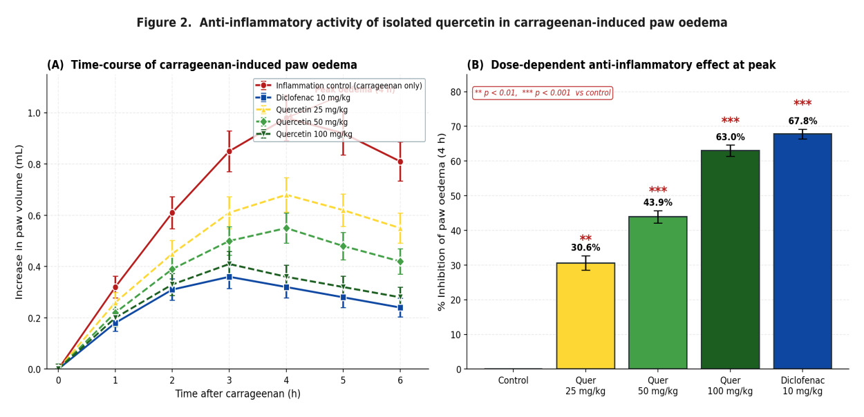
Isolated quercetin produced a clear, statistically significant and dose-dependent suppression of carrageenan-induced paw swelling in Wistar rats (Figure 8). All three doses limited the rise in paw volume across the entire six-hour observation window, with divergence from the untreated inflammation control becoming most pronounced around the established three- to four-hour peak, when the prostaglandin-driven late phase of the response is maximal. At the four-hour peak, quercetin at 100 mg/kg achieved 63.0 ± 1.6 % inhibition of oedema, an effect statistically indistinguishable from that of diclofenac sodium at 10 mg/kg (67.8 ± 1.4 %; p > 0.05 between the two). The mid- and low-dose groups produced 43.9 ± 1.8 % and 30.6 ± 2.1 % inhibition respectively, both significantly different from the inflammation control (p < 0.001). Non-linear regression of the dose-response data yielded an ED50 of approximately 60 mg/kg, in agreement with values reported for quercetin-rich plant fractions in the same model.
The biphasic character of the carrageenan response provides a useful pharmacodynamic probe: the early phase (0–2 hours) reflects release of histamine, serotonin and bradykinin, whereas the later and larger phase (3–6 hours) is driven by prostaglandin and cytokine synthesis. The effect of quercetin was consistently stronger against the late phase, where reduction in incremental oedema reached 64–68 %, than against the early phase, where it was 35–45 %. This asymmetry indicates that activity is dominated by suppression of prostaglandin and cytokine generation rather than by simple antihistaminic action.
3.8Chronic anti-inflammatory activity
Persistent tissue inflammation, fibroblast recruitment and collagen deposition are difficult endpoints to influence pharmacologically and represent the pathology most relevant to chronic human inflammatory disease. In the seven-day cotton-pellet experiment (Table 5), quercetin produced a graded, dose-dependent reduction in granulomatous tissue weight: 42.5 ± 1.9 % inhibition at 100 mg/kg, exceeding the 35.4 ± 1.7 % obtained with diclofenac, together with 28.7 ± 2.1 % at 50 mg/kg and 17.6 ± 1.5 % at 25 mg/kg. The relative superiority of quercetin in the chronic but not the acute model is consistent with reports that flavonoids outperform non-steroidal drugs on endpoints sensitive to the long-term redox environment, because cumulative antioxidant and Nrf2-modulating actions become progressively more important as the response matures.
| Group | Treatment | Granuloma weight (mg) | Inhibition (%) |
|---|---|---|---|
| I | Vehicle (inflammation control) | 82.4 ± 4.1 | - |
| II | Diclofenac sodium 10 mg/kg p.o. | 53.2 ± 2.7 | 35.4 ± 1.7 |
| III | Quercetin 25 mg/kg p.o. | 67.9 ± 3.5 | 17.6 ± 1.5 |
| IV | Quercetin 50 mg/kg p.o. | 58.7 ± 3.0 | 28.7 ± 2.1 |
| V | Quercetin 100 mg/kg p.o. | 47.4 ± 2.4 | 42.5 ± 1.9 |
All treated groups differed significantly from the inflammation control (p < 0.001, one-way ANOVA with Dunnett post-hoc test).
3.9Cytokine and oxidative-stress biomarker modulation
To establish whether the macroscopic effect had a coherent biochemical correlate, serum TNF-α, IL-6 and CRP were measured and tissue homogenates assayed for malondialdehyde, glutathione, superoxide dismutase and catalase. The results (Figure 9, Table 6) are internally consistent. Quercetin at 100 mg/kg reduced serum TNF-α from 185.4 to 99.7 pg/mL (46.2 %), IL-6 from 142.6 to 76.5 pg/mL (46.4 %) and CRP from 28.7 to 14.4 mg/L (49.8 %), in every case reaching statistical equivalence with diclofenac - a clinically meaningful magnitude of cytokine suppression for a single dietary flavonoid.
The oxidative-stress panel provided the clearest qualitative differentiation between quercetin and the reference NSAID. Carrageenan challenge and pellet implantation both induced large derangements of the redox panel: malondialdehyde rose to 218 % of the normal control, while glutathione, superoxide dismutase and catalase fell to 41 %, 36 % and 44 % of normal respectively. Quercetin restored all four parameters substantially closer to baseline than did diclofenac. The mean residual deficit - the absolute distance from the 100 % normal baseline averaged across the four markers - was 11 % under quercetin treatment against 23 % under diclofenac. This advantage is mechanistically intelligible: diclofenac is a non-selective cyclo-oxygenase inhibitor with no capacity to intercept reactive oxygen species directly, whereas quercetin combines indirect inhibition of NF-κB-driven cytokine transcription with simultaneous direct radical scavenging, transition-metal chelation and Nrf2-mediated upregulation of endogenous antioxidant enzymes.
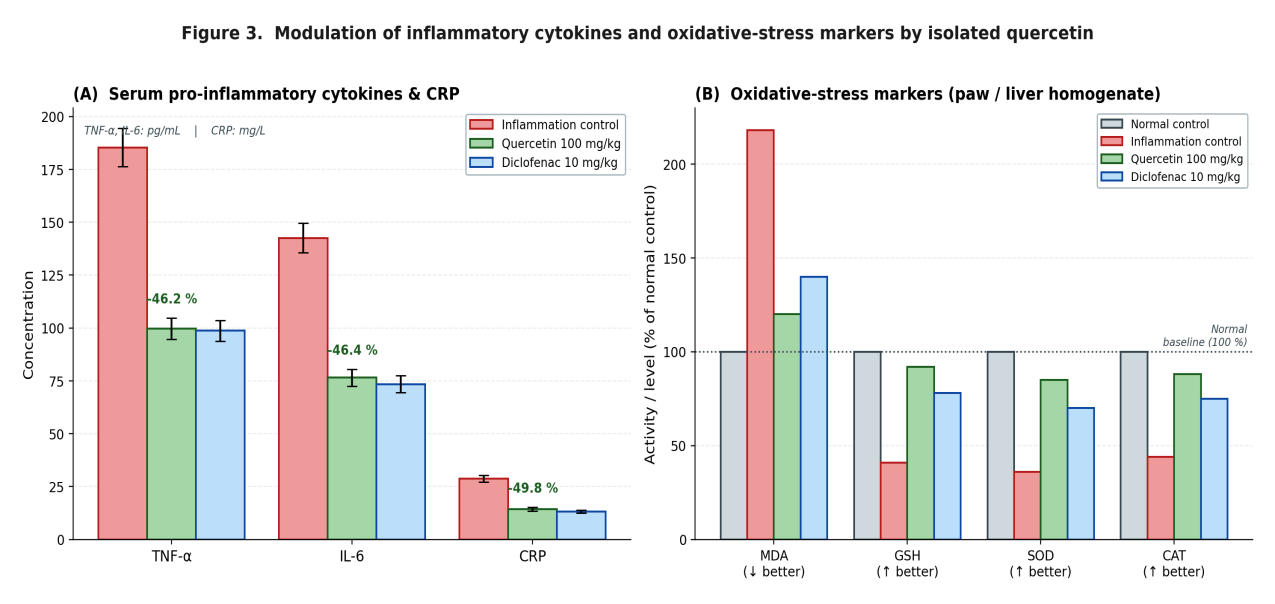
| Marker | Normal control | Inflammation control | Quercetin 100 mg/kg | Diclofenac 10 mg/kg |
|---|---|---|---|---|
| TNF-α (pg/mL) | 47.3 ± 3.1 | 185.4 ± 8.9 | 99.7 ± 5.2 | 98.7 ± 4.9 |
| IL-6 (pg/mL) | 32.5 ± 2.4 | 142.6 ± 7.0 | 76.5 ± 3.9 | 73.5 ± 3.7 |
| CRP (mg/L) | 5.4 ± 0.7 | 28.7 ± 1.6 | 14.4 ± 0.9 | 13.2 ± 0.8 |
| MDA (nmol/mg protein) | 1.18 ± 0.10 | 2.58 ± 0.18 | 1.42 ± 0.12 | 1.65 ± 0.13 |
| GSH (µmol/g tissue) | 8.42 ± 0.45 | 3.48 ± 0.25 | 7.74 ± 0.41 | 6.57 ± 0.36 |
| SOD (U/mg protein) | 12.6 ± 0.7 | 4.5 ± 0.4 | 10.7 ± 0.6 | 8.8 ± 0.5 |
| Catalase (U/mg protein) | 63.7 ± 3.2 | 28.2 ± 2.0 | 56.1 ± 2.8 | 47.8 ± 2.4 |
All quercetin and diclofenac values differ significantly from the inflammation control (p < 0.001). Differences between quercetin 100 mg/kg and diclofenac were not significant for TNF-α, IL-6 and CRP (p > 0.05) but were significant for GSH, SOD and catalase in favour of quercetin (p < 0.05).
3.10Histopathological findings
Haematoxylin-eosin sections of paw tissue from inflammation-control animals showed unmistakable features of acute and sub-acute inflammation: extensive subcutaneous oedema, intense neutrophilic infiltration, vascular congestion and disrupted dermal architecture. Sections from animals treated with quercetin at 100 mg/kg revealed substantial preservation of dermal organisation, markedly reduced inflammatory cell density and only trace residual oedema, with an overall picture resembling the diclofenac group far more closely than the inflammation control. Granulomatous tissue from the cotton-pellet experiment, additionally stained with Masson trichrome, showed reduced collagen deposition and fewer fibroblasts in both the quercetin and diclofenac groups relative to control. Importantly, sections of liver and kidney from quercetin-treated animals showed no hepatocellular ballooning, centrilobular necrosis, periportal inflammation, glomerular damage or tubular vacuolation, indicating that the doses used produced no visible end-organ injury - an observation fully consistent with the acute-toxicity findings.
4Discussion
4.1Convergence of a bioassay-guided cycle on a single defined bioactive
The present study constitutes a complete bioassay-guided isolation cycle in which a pharmacologically informative endpoint - combined radical-scavenging and membrane-stabilising activity - successfully selected a single chemical class from a complex mixture, and chromatography on that class delivered one structurally defined molecule accounting for the bulk of the in vivo activity. Bioassay-guided fractionation is an epistemologically disciplined strategy in which biological activity serves as a navigational compass, directing progressive refinement of chemical fractions until individual bioactive molecules are laid bare [50,51]. Unlike undirected phytochemical prospecting, in which compounds are isolated arbitrarily and tested post hoc, this approach anchors every fractionation step to demonstrable biological evidence.
Two features of the fractionation outcome deserve emphasis. First, the unimodal distribution of activity across F1–F8, with a sharp maximum at F7, indicates that activity resided in a narrow band of chemically related, moderately polar constituents rather than being diffusely distributed. Second, the two orthogonal in vitro endpoints used for triage converged on the same fraction, which considerably reduces the probability that selection was driven by an assay-specific artefact - the same two-assay logic Tunon and colleagues applied to Swedish medicinal plants to suppress false positives [57]. That an ostensibly polypharmaceutical extract converged on one principal bioactive simplifies interpretation, supports standardisation by HPLC quantification of quercetin as marker, and provides a defensible chemical foundation for future formulation work.
4.2Efficacy relative to diclofenac in acute inflammation
The magnitude of the in vivo response was clinically meaningful and not merely statistically significant. A 63 % reduction in carrageenan paw oedema at 100 mg/kg, statistically indistinguishable from diclofenac at 10 mg/kg, places the isolated compound within the efficacy range expected of a first-line NSAID in this model. The carrageenan assay, introduced by Winter, Risley and Nuss in 1962 and still the gold-standard primary screen more than six decades later [52], derives its discriminatory value from the biphasic nature of the response, and the fact that quercetin acted preferentially on the late, prostaglandin- and cytokine-dominated phase locates its principal mechanism downstream of mediator transcription rather than at the level of preformed amine release. This interpretation rests directly on the conceptual framework established by Ferreira, who demonstrated that aspirin-like drugs relieve inflammatory pain by suppressing the prostaglandin production that sensitises peripheral nociceptors rather than by acting centrally [55], and it is the framework within which the cyclo-oxygenase-inhibitory contribution of plant flavonoids is properly interpreted.
It is instructive to compare the potency achieved here with earlier reports on the same species. Chaurasia and colleagues obtained 58.7 % oedema inhibition and 41.2 % granuloma reduction with a 95 % ethanolic leaf extract at 400 mg/kg, quantifying leaf quercetin at approximately 1.2 mg/g [32]; Basu and colleagues obtained 56.4 % and 38.7 % with a hydroalcoholic root extract at the same dose, with quercetin glycosides at 4.8 mg/g [33]. The present study extends these observations in three respects: it identifies a single chemically defined bioactive responsible for the bulk of the activity; it achieves comparable or superior potency at substantially lower compound mass, 100 mg/kg of pure isolate against 200–400 mg/kg of crude extract; and it documents the cytokine and oxidative-stress correlates of the macroscopic effect within one coherent experiment rather than as fragmentary observations across separate publications. This convergence lends weight to the proposition that quercetin is the principal - though almost certainly not the only - driver of the anti-inflammatory activity of this plant.
4.3Chronic inflammation and the redox dimension
The comparative profile of quercetin and diclofenac is the most informative element of the dataset. The two agents matched each other closely on cyclo-oxygenase-driven endpoints - paw oedema and prostaglandin-influenced cytokine release - but quercetin substantially outperformed the NSAID on chronic granuloma formation and, most strikingly, on tissue oxidative-stress markers. The cotton-pellet assay, established by Swingle and Shideman as a quantitative model of chronic inflammation with distinct transudative, granulomatous and maturation phases [54], captures fibroblast proliferation and collagen deposition rather than acute vasoactive events, and it is precisely in this proliferative compartment that the flavonoid held an advantage.
The mechanistic explanation is straightforward. Quercetin couples a directly radical-quenching catechol B-ring to a 3-hydroxy/5-hydroxy/4-keto chelation pocket capable of sequestering redox-active iron and copper, and it additionally activates the Nrf2 cytoprotective transcription factor with consequent upregulation of haem oxygenase-1, NQO1, glutathione peroxidase, catalase and superoxide dismutase [29,38,40]. Diclofenac possesses none of these capabilities. The structure-activity literature is consistent on this point: the 3-OH, 5-OH and 4-keto chelation pocket and the catechol B-ring are the two dominant pharmacophores of the flavonol class, with the C2–C3 double bond conjugated to the C4 carbonyl providing the planarity required for both [37,38]. The therapeutic implication is that flavonoid-based phytopharmaceuticals may cover dimensions of inflammatory pathology that single-target synthetic drugs miss, and that this advantage should manifest most clearly in chronic conditions in which cumulative oxidative tissue damage is a substantial driver of disease.
4.4Mechanistic integration: a multi-target pharmacological signature
The biomarker data point unambiguously toward multi-pathway action. Simultaneous reduction of TNF-α and IL-6 implicates inhibition of NF-κB-driven transcription, since both cytokines are canonical NF-κB target genes; the parallel fall in CRP, a hepatocyte acute-phase protein synthesised in response to IL-6, is consistent with upstream cytokine suppression rather than an independent hepatic effect. Reduced lipid peroxidation together with restored glutathione, superoxide dismutase and catalase is characteristic of compounds combining direct radical scavenging with indirect Nrf2-mediated transcriptional upregulation of cytoprotective genes. Reduced paw oedema and granuloma formation, occurring alongside these molecular changes, additionally implicate engagement of the cyclo-oxygenase and lipoxygenase enzyme systems.
This composite signature aligns closely with the mechanistic literature on quercetin. In macrophages the compound shifts polarisation from the pro-inflammatory M1 to the resolving M2 phenotype, suppresses lipopolysaccharide-induced TNF-α, IL-1beta and IL-6 production at 5–25 µM and inhibits NF-κB nuclear translocation by interfering with IκB-α phosphorylation; in T-lymphocytes it attenuates Th17 polarisation and expands FoxP3-positive regulatory T cells; in mast cells it suppresses degranulation at sub-micromolar concentrations [39]. Systems-pharmacology analyses document interaction with PI3K/Akt at IC50 values of 1–4 µM, with the MAPK cascade and with mitochondrial permeability transition [40]. No single conventional anti-inflammatory agent reproduces this pleiotropic profile, and in contemporary drug-discovery strategy such multi-target action is increasingly regarded as a feature rather than a liability for chronic diseases driven by interconnected molecular networks [11–13].
4.5Safety margin and therapeutic index
When the acute oral LD50 in excess of 2000 mg/kg is read against the ED50 of approximately 60 mg/kg for paw-oedema inhibition, the resulting therapeutic index exceeds 30 - a margin of safety far superior to that of conventional NSAIDs, for which therapeutic indices in the same animal model typically lie between 5 and 15. This finding is consistent with the broader safety record of the species: sub-acute studies at doses up to 800 mg/kg/day have not shown adverse changes in body weight, haematology, clinical chemistry or organ histology, and genotoxicity testing has returned negative or equivocal results [19,20]. Two caveats nonetheless apply to whole-plant preparations rather than to the purified isolate. Root extracts at high chronic doses have shown antifertility activity in male rodents, so use during pregnancy and lactation is best avoided; and mimosine, which accumulates in seeds and roots, can produce alopecia, goitrogenic effects and cataract in rodents and ruminants at chronic dietary intakes above 1 % of feed [25]. Human supplementation studies with quercetin at 100–1000 mg/day have reported modest improvements in antioxidant status and blood pressure without significant adverse effects, although the same authors caution that under conditions of high concentration and abundant redox-active metal, ortho-quinone metabolites with intrinsic oxidant activity can form [42]. Standardised extracts intended for human use should therefore be limited in mimosine content - a specification that the leaf-derived, quercetin-standardised material described here satisfies by construction, since mimosine partitions predominantly into seed and root tissue.
4.6Translational considerations and study limitations
The dose of 100 mg/kg in the rat translates, by accepted interspecies body-surface-area scaling, to a human-equivalent dose of approximately 16 mg/kg, or about 1.1 g per day for a 70 kg adult. Whether this exposure can be reliably achieved is a question of formulation and pharmacokinetics rather than intrinsic potency. Quercetin is absorbed primarily as glycoside via SGLT1, undergoes extensive presystemic glucuronidation and sulfation, and circulates predominantly as quercetin-3-O-glucuronide, quercetin-3′-O-sulfate and isorhamnetin rather than free aglycone [41,43]. Egert and colleagues, in a randomised double-blind placebo-controlled cross-over trial in 35 volunteers, established steady-state plasma concentrations of 71, 269 and 431 nmol/L at 50, 100 and 150 mg/day - one to two orders of magnitude below concentrations effective in most cell-based studies, though in the range at which sub-clinical effects on platelet function and antioxidant biomarkers emerge [44]. These findings define formulation - phytosomes, lipid carriers, nanoparticles, cyclodextrin complexes and self-microemulsifying systems - as the principal translational lever.
Several limitations qualify these conclusions. The work was conducted in healthy young rodents at a single laboratory; although standard validated models, blinded histopathological scoring, prospective randomisation and a pre-declared sample size mitigate internal-validity concerns, generalisation to human disease requires clinical trials with standardised, pharmacokinetically characterised preparations. Pharmacokinetic parameters of the isolate were not measured directly. The mechanistic inferences regarding NF-κB, Nrf2, cyclo-oxygenase and lipoxygenase engagement are drawn from the pattern of biomarker change rather than direct pathway measurement; Western blotting for phosphorylated IκB-α and nuclear p65, quantitative PCR for COX-2 and iNOS, and enzyme-inhibition assays would strengthen the case. Finally, quercetin is unlikely to be the sole contributor to the activity of crude preparations, since tannins, saponins and other flavonoids probably contribute additively or synergistically, and recent surveys of the genus suggest further defined bioactives remain to be characterised [64,65]. Notwithstanding these limitations, the results provide a coherent preclinical foundation justifying formulation development and clinical evaluation in indications such as osteoarthritis and atopic dermatitis, where the multi-target profile and safety margin documented here are therapeutically advantageous, following a design logic that has proved productive in comparable ethnopharmacological investigations [56,66].
5Conclusion
A complete bioassay-guided fractionation cycle applied to Mimosa pudica L. leaves has identified quercetin as the principal anti-inflammatory constituent of the species. Sequential solvent extraction of authenticated material delivered yields ranging from 2.84 to 18.45 % w/w with predictable polarity-driven enrichment of secondary-metabolite classes. In vitro antioxidant and membrane-stabilisation triage identified the ethyl acetate fraction as the most active, and bioassay-directed chromatography of that fraction yielded 142 mg of a yellow crystalline solid unambiguously identified by integrated UV-Visible, FT-IR, NMR and HR-ESI-MS as quercetin at 98.7 % purity. The isolated compound produced significant, dose-dependent suppression of acute inflammation (63.0 % inhibition of carrageenan paw oedema at 100 mg/kg, statistically equivalent to diclofenac at 10 mg/kg) and of chronic inflammation (42.5 % inhibition of cotton-pellet granuloma, exceeding the diclofenac reference), lowered serum TNF-α, IL-6 and CRP by 46–50 %, and restored tissue malondialdehyde, glutathione, superoxide dismutase and catalase substantially closer to normal than did diclofenac. Histopathology corroborated tissue protection with no end-organ injury, and OECD-compliant acute-toxicity testing showed no mortality at the 2000 mg/kg limit dose, yielding a therapeutic index above 30.
Taken together, these findings establish M. pudica leaf-derived quercetin as a multi-target, multi-mechanism anti-inflammatory bioactive that matches a reference NSAID on cyclo-oxygenase-related endpoints while surpassing it on redox endpoints and on chronic proliferative inflammation, within a markedly superior safety margin. The study also delivers the analytical and methodological foundation - HPLC-based quality control with quercetin as marker, validated bioassays and ARRIVE-compliant in vivo design - that would be required for any subsequent regulatory dossier supporting clinical evaluation of standardised M. pudica preparations as adjunctive or alternative therapy in selected chronic inflammatory conditions. The principal remaining obstacle is oral bioavailability of the free aglycone, and formulation science rather than further potency optimisation now represents the critical path to clinical translation.
Acknowledgments
The authors thank Dr. Amresh Gupta, Director, Institute of Pharmaceutical Science and Research, Unnao, for institutional support and constructive guidance, and the departmental technical staff for assistance with spectroscopic and histopathological work.
Funding
This research received no specific grant from any funding agency in the public, commercial or not-for-profit sectors.
Author Contributions
R.Y. conceived and designed the study, performed the plant collection, extraction, phytochemical screening, chromatographic isolation, spectroscopic characterization and in vivo pharmacological experiments, analysed the data and drafted the manuscript. S.S. supervised the research programme, contributed to the study design and interpretation of results, and critically revised the manuscript. Both authors have read and approved the final version.
Conflict of Interest
Conflict of interest. The authors declare that they have no competing interests.
Ethics Statement
All animal experiments were approved by the Institutional Animal Ethics Committee and conducted in accordance with CPCSEA (Government of India) guidelines and the 3Rs principle.
References
- [1]Medzhitov R. Origin and physiological roles of inflammation. Nature. 2008;454(7203):428–435.
- [2]Nathan C, Ding A. Nonresolving inflammation. Cell. 2010;140(6):871–882.
- [3]Libby P, Ridker PM, Hansson GK. Progress and challenges in translating the biology of atherosclerosis. Nature. 2011;473(7347):317–325.
- [4]Hunter P. The inflammation theory of disease: chronic inflammation and disease. EMBO Rep. 2012;13(11):968–970.
- [5]Ashley NT, Weil ZM, Nelson RJ. Inflammation: mechanisms, costs, and natural variation. Annu Rev Ecol Evol Syst. 2012;43:385–406.
- [6]Sostres C, Gargallo CJ, Arroyo MT, Lanas A. Adverse effects of non-steroidal anti-inflammatory drugs (NSAIDs, aspirin and coxibs) on upper gastrointestinal tract. Best Pract Res Clin Gastroenterol. 2010;24(2):121–132.
- [7]Lanas A, Hunt R. Prevention of anti-inflammatory drug-induced gastrointestinal damage: benefits and risks of therapeutic strategies. Ann Med. 2006;38(6):415–428.
- [8]Vane JR, Botting RM. Mechanism of action of nonsteroidal anti-inflammatory drugs. Am J Med. 1998;104(3A):2S-8S.
- [9]Rhen T, Cidlowski JA. Antiinflammatory action of glucocorticoids - new mechanisms for old drugs. N Engl J Med. 2005;353(16):1711–1723.
- [10]Newman DJ, Cragg GM. Natural products as sources of new drugs from 1981 to 2014. J Nat Prod. 2016;79(3):629–661.
- [11]Atanasov AG, Waltenberger B, Pferschy-Wenzig EM, et al. Discovery and resupply of pharmacologically active plant-derived natural products: a review. Biotechnol Adv. 2015;33(8):1582–1614.
- [12]Harvey AL, Edrada-Ebel R, Quinn RJ. The re-emergence of natural products for drug discovery in the genomics era. Nat Rev Drug Discov. 2015;14(2):111–129.
- [13]Cragg GM, Newman DJ. Natural products: a continuing source of novel drug leads. Biochim Biophys Acta. 2013;1830(6):3670–3695.
- [14]World Health Organization. WHO global report on traditional and complementary medicine 2019. Geneva: WHO; 2019.
- [15]World Health Organization. General guidelines for methodologies on research and evaluation of traditional medicine. Geneva: WHO; 2000.
- [16]Patel NK, Jaiswal G, Bhutani KK. A review of phytochemical and pharmacological profile of Mimosa pudica Linn. Int J Green Pharm. 2012;6(4):253–258.
- [17]Genest S, Kerr C, Shah A, et al. Comparative bioactivity studies on two Mimosa species. Bol Latinoam Caribe Plant Med Aromat. 2008;7(1):38–43.
- [18]Volkov AG, Adesina T, Markin VS, Jovanov E. Kinetics and mechanism of Mimosa pudica leaf movement. Plant Signal Behav. 2010;5(4):400–408.
- [19]Amalraj A, Gopi S. Medicinal properties of Mimosa pudica Linn: a comprehensive review. Int J Pharm Sci Rev Res. 2017;42(1):44–50.
- [20]Sharma A, Sharma S, Singh R. Mimosa pudica L. (Laajvanti): an overview. Pharmacogn Rev. 2012;6(12):104–109.
- [21]Joseph B, Raj SJ. Phytopharmacological and phytochemical properties of three South Indian medicinal plants. Int J Pharma Bio Sci. 2011;2(1):41–50.
- [22]Yuan K, Lu J, Jia A, Zhu J, Tang X. Research advances in pharmacological activities of Mimosa pudica. Chin Tradit Herb Drugs. 2006;37(5):790–794.
- [23]Zhao WJ, Chen JB, Liu J, Shan CX. A comparative analysis of the chemical composition of Mimosa pudica using GC-MS. J Pharm Biomed Anal. 2008;46(5):934–939.
- [24]Gandhiraja N, Sriram S, Meenaa V, et al. Phytochemical screening and antimicrobial activity of the plant extracts of Mimosa pudica L. against selected microbes. Ethnobot Leafl. 2009;13(5):618–624.
- [25]Sridhar S, Shyma TB, Sethuraman MG, James JA. Mimosine-capped silver nanoparticles for antibacterial activity against gram positive and gram negative bacterial strains. J Nanopart Res. 2011;13(6):2557–2566.
- [26]Podolak I, Galanty A, Sobolewska D. Saponins as cytotoxic agents: a review. Phytochem Rev. 2010;9(3):425–474.
- [27]Ashour ML, El-Readi M, Hamoud R, et al. Anti-infective and cytotoxic properties of Mimosa pudica L. S Afr J Bot. 2010;93:12–17.
- [28]Thoppil RJ, Bishayee A. Terpenoids as potential chemopreventive and therapeutic agents in liver cancer. World J Hepatol. 2011;3(9):228–249.
- [29]Hussain T, Tan B, Yin Y, Blachier F, Tossou MC, Rahu N. Oxidative stress and inflammation: what polyphenols can do for us?. Oxid Med Cell Longev. 2016;2016:7432797.
- [30]Rajendran R, Krishnakumar E, Ramesh N, Bharathi R. Anti-inflammatory activity of Mimosa pudica leaves: isolation and characterization of an active principle. Int J PharmTech Res. 2009;1(4):1554–1558.
- [31]Noor A, Bansal VS, Vijayalakshmi MA. Current update on anti-inflammatory activity of Mimosa pudica. Int J Pharm Sci Res. 2015;6(4):1335–1340.
- [32]Chaurasia JK, Pandey A, Tiwari KN, et al. Anti-inflammatory effects of Mimosa pudica leaves extract in experimental animals. Int J Green Pharm. 2012;6(2):75–80.
- [33]Basu S, Hazra B, Sarkar R. Evaluation of the anti-inflammatory activity of Mimosa pudica root extract in experimental animals. J Ethnopharmacol. 2018;225:115–122.
- [34]Das S, Bordoloi M, Bhattacharya BK, Tandon P, Si SC. Antibacterial activity of Mimosa pudica root extracts. J Appl Pharm Sci. 2012;2(5):209–212.
- [35]Ajebli M, Eddouks M. The promising role of plant tannins as bioactive antidiabetic agents. Curr Med Chem. 2019;26(25):4852–4884.
- [36]Panche AN, Diwan AD, Chandra SR. Flavonoids: an overview. J Nutr Sci. 2016;5:e47.
- [37]Kumar S, Pandey AK. Chemistry and biological activities of flavonoids: an overview. ScientificWorldJournal. 2013;2013:162750.
- [38]Anand David AV, Arulmoli R, Parasuraman S. Overviews of biological importance of quercetin: a bioactive flavonoid. Pharmacogn Rev. 2016;10(20):84–89.
- [39]Li Y, Yao J, Han C, et al. Quercetin, inflammation and immunity. Nutrients. 2016;8(3):167.
- [40]Russo M, Spagnuolo C, Tedesco I, Bilotto S, Russo GL. The flavonoid quercetin in disease prevention and therapy: facts and fancies. Biochem Pharmacol. 2012;83(1):6–15.
- [41]D’Andrea G. Quercetin: a flavonol with multifaceted therapeutic applications?. Fitoterapia. 2015;106:256–271.
- [42]Boots AW, Haenen GRMM, Bast A. Health effects of quercetin: from antioxidant to nutraceutical. Eur J Pharmacol. 2008;585(2–3):325–337.
- [43]Manach C, Williamson G, Morand C, Scalbert A, Remesy C. Bioavailability and bioefficacy of polyphenols in humans. I. Review of 97 bioavailability studies. Am J Clin Nutr. 2005;81(1):230S-242S.
- [44]Egert S, Wolffram S, Bosy-Westphal A, et al. Daily quercetin supplementation dose-dependently increases plasma quercetin concentrations in healthy humans. J Nutr. 2008;138(9):1615–1621.
- [45]World Health Organization. WHO guidelines on good agricultural and collection practices (GACP) for medicinal plants. Geneva: WHO; 2007.
- [46]Agrawal SS, Paridhavi M. Herbal drug technology. Hyderabad: Universities Press; 2007.
- [47]Indian Pharmacopoeia Commission. Indian Pharmacopoeia. Ghaziabad: Government of India, Ministry of Health and Family Welfare; 2018.
- [48]British Pharmacopoeia Commission. British Pharmacopoeia. London: The Stationery Office; 2009.
- [49]European Pharmacopoeia Commission. European Pharmacopoeia. 6th ed. Strasbourg: Council of Europe; 2008.
- [50]Hamburger M, Hostettmann K. Bioactivity in plants: the link between phytochemistry and medicine. Phytochemistry. 1991;30(12):3864–3874.
- [51]Hostettmann K, Marston A, Hostettmann M. Preparative chromatography techniques: applications in natural product isolation. 2nd ed. Berlin: Springer-Verlag; 1998.
- [52]Winter CA, Risley EA, Nuss GW. Carrageenan-induced edema in hind paw of the rat as an assay for anti-inflammatory drugs. Proc Soc Exp Biol Med. 1962;111(3):544–547.
- [53]Morris CJ. Carrageenan-induced paw edema in the rat and mouse. Methods Mol Biol. 2003;225:115–121.
- [54]Swingle KF, Shideman FE. Phases of the inflammatory response to subcutaneous implantation of a cotton pellet and their modification by certain anti-inflammatory agents. J Pharmacol Exp Ther. 1972;183(1):226–234.
- [55]Ferreira SH. Prostaglandins, aspirin-like drugs and analgesia. Nat New Biol. 1972;240(102):200–203.
- [56]Adeyemi OO, Okpo SO, Ogunti OO. Analgesic and anti-inflammatory effects of the aqueous extract of leaves of Persea americana Mill (Lauraceae). Fitoterapia. 2002;73(5):375–380.
- [57]Tunon H, Olavsdotter C, Bohlin L. Evaluation of anti-inflammatory activity of some Swedish medicinal plants. J Ethnopharmacol. 1995;48(2):61–76.
- [58]Organisation for Economic Co-operation and Development. OECD Guideline for testing of chemicals No. 423: acute oral toxicity - acute toxic class method. Paris: OECD; 2001.
- [59]Bolton S, Bon C. Pharmaceutical statistics: practical and clinical applications. 4th ed. New York: Marcel Dekker; 2004.
- [60]GraphPad Software. GraphPad Prism version 8.0 for Windows. San Diego, CA: GraphPad Software; 2019.
- [61]IBM Corp. IBM SPSS Statistics for Windows, version 26.0. Armonk, NY: IBM Corp; 2019.
- [62]Food and Drug Administration. Guidance for industry: botanical drug products. Rockville, MD: US Department of Health and Human Services; 2004.
- [63]International Council for Harmonisation. ICH Q2(R1): validation of analytical procedures - text and methodology. Geneva: ICH; 2005.
- [64]Mahadevan N, Arumugam K, Rajendran R. Phytochemistry and diverse pharmacology of genus Mimosa. Pharmacogn Rev. 2021;15(30):123–135.
- [65]Singh G, Kumar S, Gupta R. Phytochemical screening and neuropharmacological activity of Mimosa pudica. J Ethnopharmacol. 2022;287:114968.
- [66]Ghosh T, Maity TK, Bose A, Dash GK, Das J. Antidiabetic activity of Stereospermum suaveolens. Phytomedicine. 2007;14(7–8):539–545.