In Vitro Antidiabetic Activity of Medicinal Plants
Comparative In Vitro Evaluation of the Antidiabetic Potential of Morinda lucida and Ficus capensis Leaf Extracts
Department of Biochemistry, Bingham University, Karu, Nasarawa State, Nigeria
Published October 2026
Abstract
Oxidative stress and rapid postprandial hyperglycaemia are central features of diabetes mellitus, and plant extracts that scavenge free radicals and inhibit the carbohydrate-digesting enzymes α-amylase and α-glucosidase are of interest as sources of antidiabetic agents. Morinda lucida and Ficus capensis are tropical African medicinal plants with reported antidiabetic uses, but their in vitro activities have not been compared directly under the same conditions. Ethanolic leaf extracts of both plants were evaluated for total phenolic content (TPC; Folin–Ciocalteu), total flavonoid content (TFC; aluminium chloride), DPPH radical-scavenging activity (62.5–1000 µg/mL), ferric reducing antioxidant power (FRAP), and α-amylase and α-glucosidase inhibition, with acarbose as the reference inhibitor. IC50 values were estimated by nonlinear regression. F. capensis had higher TPC (≈240 versus ≈85 µg GAE/mg) and TFC (≈123 versus ≈101 µg QE/mg) than M. lucida. DPPH scavenging at 1000 µg/mL was 77.98 ± 0.33% for F. capensis versus 30.37 ± 0.62% for M. lucida, and FRAP was ≈95 versus ≈20 µg TE/mg. F. capensis showed stronger α-amylase inhibition (IC50 ≈110 versus ≈500 µg/mL; acarbose ≈90 µg/mL) and a slightly lower α-glucosidase IC50 (≈230 versus ≈250 µg/mL; acarbose ≈100 µg/mL). Under the conditions tested, the ethanolic leaf extract of F. capensis showed the stronger antioxidant and carbohydrate-digestive-enzyme inhibitory profile, particularly against α-amylase, whereas M. lucida showed weaker but measurable activity. These in vitro findings support further bioassay-guided and in vivo investigation of both plants as sources of antidiabetic constituents.
Keywords: α-amylase; α-glucosidase; antioxidant activity; Ficus capensis; Morinda lucida
1Introduction
Diabetes mellitus is a heterogeneous metabolic disorder characterized by impaired insulin secretion, impaired insulin action, or both, resulting in persistent hyperglycaemia and disturbances in carbohydrate, lipid and protein metabolism. The global burden of diabetes continues to increase, with the International Diabetes Federation estimating 537 million adults living with diabetes in 2021 and projecting approximately 783 million by 2045 [1]. The disease is accompanied by progressive vascular and tissue complications and remains an important public-health challenge in Nigeria and other low- and middle-income settings [2], [3].
Hyperglycaemia is closely linked to oxidative stress. Excess reactive oxygen species can impair insulin signaling and damage pancreatic β-cells, thereby contributing to insulin resistance, β-cell dysfunction and diabetic complications [4]–[6]. Antioxidant compounds that scavenge free radicals or reduce oxidized species are therefore of interest in diabetes research. In addition, delaying carbohydrate digestion by inhibiting pancreatic α-amylase, which hydrolyses starch to oligosaccharides, and intestinal α-glucosidase, which releases absorbable glucose, is an established approach to blunting postprandial hyperglycaemia.
Although conventional antidiabetic medicines are effective, long-term management can be limited by cost, access, adverse effects and treatment failure. Consequently, medicinal plants remain important sources of candidate antidiabetic agents, particularly in settings where traditional medicine is widely used [7]–[9]. Plant phenolics, flavonoids and other secondary metabolites may influence glucose metabolism through antioxidant and carbohydrate-digestive-enzyme inhibitory mechanisms [10]–[14]. Simple in vitro assays of phenolic content, radical scavenging and enzyme inhibition provide a rapid and inexpensive way to compare candidate extracts before animal studies are undertaken.
Morinda lucida Benth. (Rubiaceae) is an African medicinal plant with documented ethnomedicinal use and reported antioxidant and hypoglycaemic properties [15], [16]. Its phenolic profile and biological activity have been shown to depend on the extraction method used [17], and leaf preparations have improved glycaemic status and selected metabolic indices in alloxan-induced diabetic rats [18]–[20]. Ficus capensis Thunb. (Moraceae), commonly known as bush fig, is another tropical African plant used in traditional medicine and as a food plant, and glucose-lowering effects of F. capensis-containing preparations have been reported in diabetic rats [21], [22].
Despite evidence supporting the antidiabetic potential of both plants, direct comparison of their antioxidant and carbohydrate-digestive-enzyme inhibitory activities under the same experimental conditions is limited. The present study therefore compared the total phenolic and flavonoid contents, antioxidant activities (DPPH and FRAP) and α-amylase and α-glucosidase inhibitory activities of ethanolic M. lucida and F. capensis leaf extracts in vitro, using acarbose as the reference inhibitor, to determine which extract shows the stronger antidiabetic potential in these assays. The in vivo glycaemic, lipid-modulating, molecular and histological effects of the same extracts in alloxan-induced diabetic rats are reported in a companion manuscript.
2Materials and methods
2.1Plant materials and extraction
Mature, fresh and disease-free leaves of M. lucida and F. capensis were collected, washed, shade-dried at room temperature to constant weight, pulverized and extracted with ethanol. The plants were authenticated at the National Institute for Pharmaceutical Research and Development (NIPRD), Abuja. Extraction and subsequent analyses were conducted in specialized laboratories in Nigeria as described in the dissertation record. The ethanolic extracts were used for all assays described below.
2.2Determination of total phenolic content
Total phenolic content was determined by the Folin–Ciocalteu colorimetric method. Extract solution (1 mg/mL) was reacted with 10% Folin–Ciocalteu reagent and 7.5% sodium carbonate, incubated in the dark and read at 750 nm. Gallic acid was used for calibration and results were expressed as µg gallic acid equivalents per mg extract (µg GAE/mg). Determinations were performed in triplicate.
2.3Determination of total flavonoid content
Total flavonoid content was determined using the aluminium chloride colorimetric method with quercetin as the reference standard, with absorbance measured at 415 nm and results expressed as µg quercetin equivalents per mg extract (µg QE/mg). Determinations were performed in triplicate.
2.4DPPH radical-scavenging assay
DPPH radical-scavenging activity was assessed using extract concentrations of 62.5–1000 µg/mL [23]. Following incubation for 30 min in the dark, absorbance was measured at 517 nm. Percentage scavenging was calculated as [(Absorbance control − Absorbance sample)/Absorbance control] × 100.
2.5Ferric reducing antioxidant power (FRAP) assay
FRAP was assessed using a microplate procedure with a freshly prepared acetate buffer/TPTZ/ferric chloride reagent and absorbance at 593 nm [24]. Trolox was used as the calibration standard and activity was expressed as µg Trolox equivalents per mg extract (µg TE/mg).
2.6α-amylase inhibition assay
α-Amylase inhibition was assessed using a starch hydrolysis assay with porcine pancreatic α-amylase. Extracts were tested over a concentration range and reaction products were quantified spectrophotometrically. Acarbose was used as the reference inhibitor. Percentage inhibition was calculated from control and sample absorbances, and IC50 values were estimated from dose–response relationships using nonlinear regression.
2.7α-glucosidase inhibition assay
α-Glucosidase inhibition was determined using p-nitrophenyl-α-D-glucopyranoside as substrate, with absorbance measured at 405 nm [25]. Extracts were tested over a concentration range with acarbose as the reference inhibitor. Percentage inhibition was calculated from control and sample absorbances, and IC50 values were estimated from dose–response relationships using nonlinear regression.
2.8Statistical analysis
Results were expressed as mean ± standard deviation of triplicate determinations. Differences among groups were evaluated using one-way analysis of variance followed by Tukey’s HSD post-hoc test where appropriate; independent-samples t-tests were used for the two-group comparisons. Statistical significance was accepted at p < 0.05. Dose–response IC50 values were obtained by nonlinear regression.
3Results
3.1Total phenolic and flavonoid contents
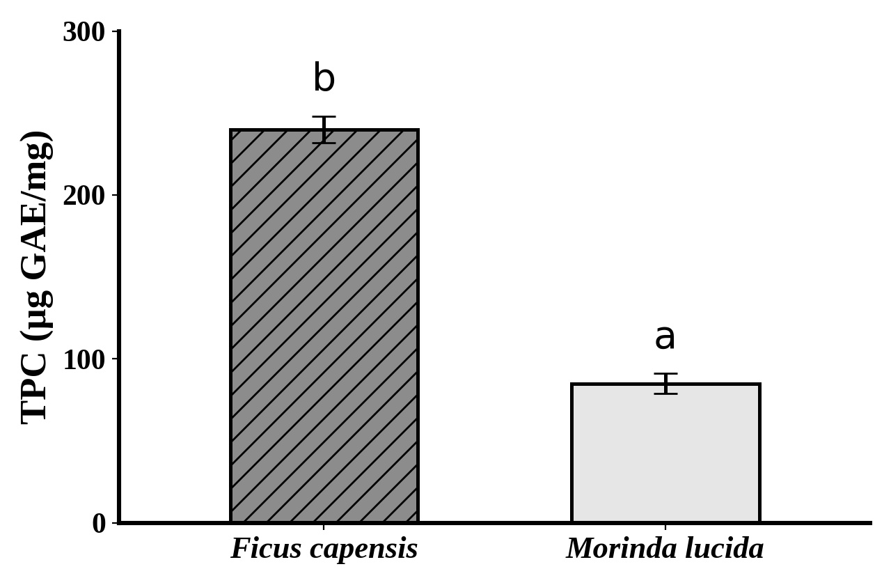
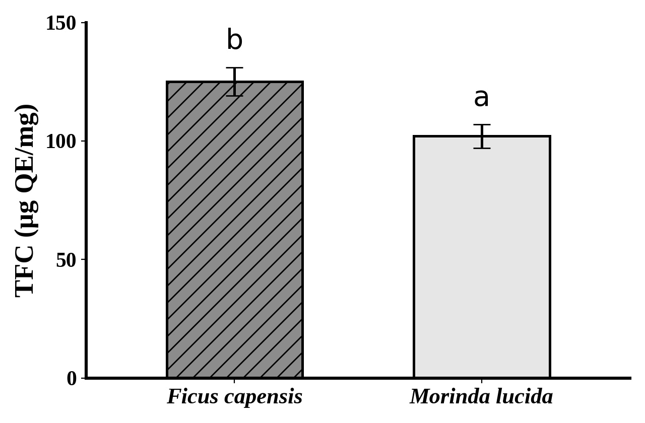
F. capensis contained substantially more total phenolic material than M. lucida, with values of approximately 240 and 85 µg GAE/mg, respectively (Figure 1). Total flavonoid content was also higher in F. capensis (approximately 123 µg QE/mg) than in M. lucida (approximately 101 µg QE/mg) (Figure 2). The differences between the extracts were statistically significant (p < 0.05).
3.2DPPH radical-scavenging activity
F. capensis exhibited greater DPPH radical-scavenging activity than M. lucida at 125–1000 µg/mL, whereas the extracts were statistically similar at 62.5 µg/mL (Table 1). At 1000 µg/mL, F. capensis produced 77.98 ± 0.33% inhibition compared with 30.37 ± 0.62% for M. lucida. The activity increased with concentration in both extracts.
| Conc. (µg/mL) | F. capensis (% inhibition) | M. lucida (% inhibition) |
|---|---|---|
| 1000 | 77.98 ± 0.33a | 30.37 ± 0.62b |
| 500 | 60.19 ± 0.35a | 17.95 ± 0.97b |
| 250 | 34.63 ± 0.28a | 15.83 ± 0.69b |
| 125 | 23.39 ± 0.39a | 13.08 ± 0.65b |
| 62.5 | 9.24 ± 0.20a | 9.67 ± 0.75a |
| 0 | 0.00 ± 0.00a | 0.00 ± 0.00a |
Note: Values are mean ± SD of triplicate determinations. Different superscript letters within a row indicate significant difference at p < 0.05.
3.3Ferric reducing antioxidant power
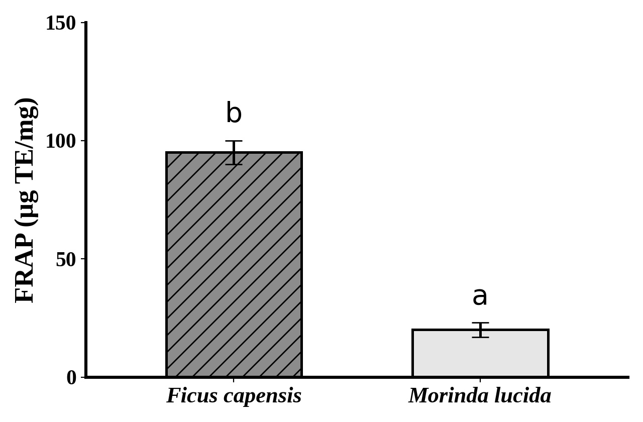
F. capensis showed markedly greater ferric-reducing capacity than M. lucida, with approximate values of 95 and 20 µg TE/mg, respectively (Figure 3).
3.4α-glucosidase inhibition
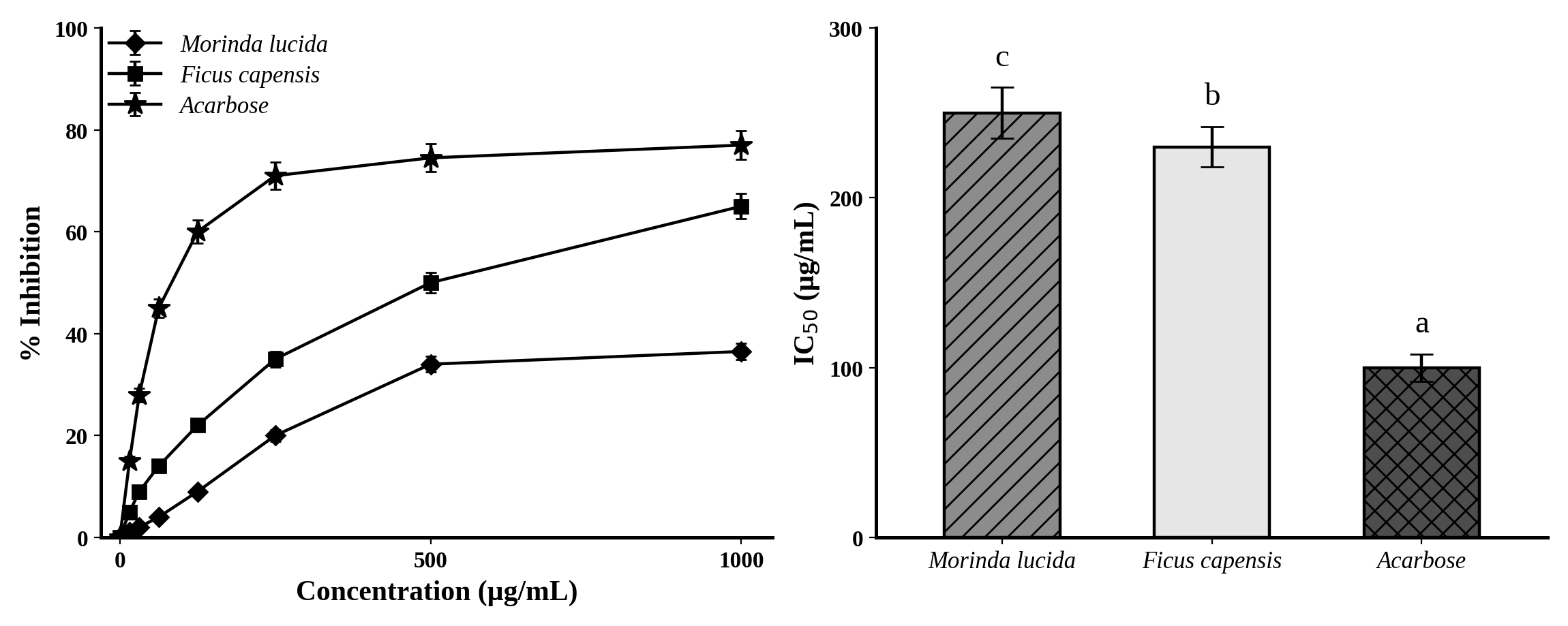
Both extracts inhibited α-glucosidase in a concentration-dependent manner (Figure 4). F. capensis produced approximately 65% inhibition at 1000 µg/mL compared with approximately 36% for M. lucida, while acarbose produced approximately 77%. The corresponding IC50 values were approximately 230 µg/mL for F. capensis, 250 µg/mL for M. lucida and 100 µg/mL for acarbose.
3.5α-amylase inhibition
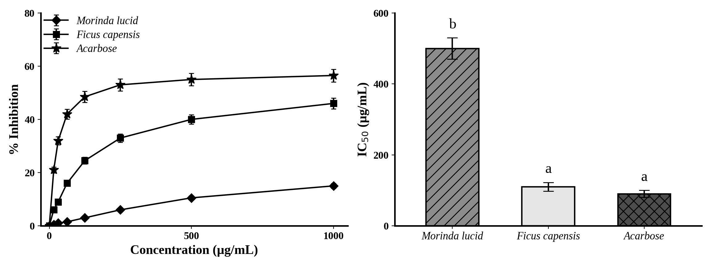
F. capensis demonstrated considerably stronger α-amylase inhibition than M. lucida (Figure 5). At 1000 µg/mL, inhibition was approximately 46% for F. capensis and 15% for M. lucida, compared with approximately 56% for acarbose. IC50 values were approximately 110 µg/mL for F. capensis, 500 µg/mL for M. lucida and 90 µg/mL for acarbose.
4Discussion
4.1Phenolic and flavonoid contents
The substantially higher TPC and TFC observed in F. capensis compared with M. lucida indicate that the two ethanolic extracts were chemically distinct under the conditions of the present study. Phenolic compounds and flavonoids can contribute to antioxidant and antidiabetic activities through electron- or hydrogen-donation reactions and through interactions with metabolic enzymes. Recent evidence coninues to support the relevance of phenolic-rich plant matrices in diabetes research, while also emphasizing that total-content assays do not identify the individual molecules responsible for activity [13], [26]. The phenolic profile and biological activity of M. lucida extracts have also been shown to depend on the extraction method [17], so the values reported here should be interpreted as specific to the solvent and conditions employed.
4.2Antioxidant activity
The antioxidant results showed strong convergence. F. capensis outperformed M. lucida in DPPH activity at most concentrations and had substantially greater FRAP reducing capacity. The parallel superiority across TPC, DPPH and FRAP suggests that the stronger antioxidant activity of F. capensis is related, at least in part, to a richer or more effective mixture of reducing constituents. The antioxidant activity of M. lucida leaf extracts has been described previously [16], and the lower activity observed here may reflect the extraction solvent and conditions. Because oxidative stress contributes to β-cell injury and insulin resistance [4], [6], antioxidant capacity is a relevant property for antidiabetic candidates. However, DPPH and FRAP are chemical assays and should not be interpreted as direct measurements of antioxidant effects in a biological system.
4.3Carbohydrate-digestive-enzyme inhibition
Both extracts inhibited α-amylase and α-glucosidase in a concentration-dependent manner, supporting their potential to interfere with carbohydrate digestion. F. capensis showed a particularly clear advantage against α-amylase, with an IC50 of approximately 110 µg/mL compared with approximately 500 µg/mL for M. lucida, a difference of roughly four- to five-fold. Its α-glucosidase IC50 was also slightly lower than that of M. lucida. These results are consistent with contemporary evidence that flavonoids and other plant constituents can inhibit both carbohydrate-digestive enzymes, although the magnitude of inhibition varies according to plant species and chemical composition [27]–[29].
The fact that acarbose remained more potent than the crude extracts in the α-glucosidase assay (IC50 approximately 100 µg/mL versus 230–250 µg/mL) is important. A purified pharmaceutical inhibitor cannot be considered equivalent to a chemically complex crude extract on the basis of an in vitro IC50 alone. Conversely, the relatively close α-amylase IC50 values of F. capensis (approximately 110 µg/mL) and acarbose (approximately 90 µg/mL) suggest that F. capensis contains constituents capable of substantial enzyme inhibition under the assay conditions. Enzyme-kinetic studies using purified fractions would be needed to determine the precise mode of inhibition.
4.4Overall in vitro comparison
Taken together, F. capensis demonstrated the broader and stronger in vitro profile, with higher TPC and TFC, stronger DPPH and FRAP activities, stronger α-amylase inhibition and somewhat stronger α-glucosidase inhibition. M. lucida showed measurable but weaker activity in every assay. The study does not establish that the higher phenolic content caused the stronger enzyme inhibition, because total-content assays do not identify the active molecules, and the activities of crude extracts cannot be extrapolated directly to effects in living organisms, where absorption, metabolism and bioavailability influence activity. The in vitro findings are therefore best regarded as a screening-level indication of antidiabetic potential that justifies, and should be tested by, in vivo investigation, which is reported separately for the same extracts.
5Conclusion
Ethanolic leaf extracts of Morinda lucida and Ficus capensis demonstrated measurable antioxidant and carbohydrate-digestive-enzyme inhibitory activities in vitro. Ficus capensis exhibited higher phenolic and flavonoid contents, stronger DPPH-scavenging and ferric-reducing activities, considerably stronger α-amylase inhibition and slightly stronger α-glucosidase inhibition than Morinda lucida, with α-amylase inhibition approaching that of acarbose. These results indicate that F. capensis has the stronger antidiabetic potential in these assays, support both plants as sources of bioactive constituents, and justify bioassay-guided fractionation and in vivo evaluation. The evidence is in vitro only and does not demonstrate antidiabetic efficacy in animals or humans.
5.1Limitations
The principal limitations were the use of crude ethanolic extracts prepared with a single solvent, the chemical (non-cellular) nature of the antioxidant assays, the use of total phenolic and flavonoid assays that do not identify individual compounds, the absence of enzyme-kinetic analysis to define the mode of inhibition, and the lack of chemical fingerprinting and standardization of the extracts. The assays do not account for absorption, metabolism or bioavailability.
5.2Recommendations
- 1. Use HPLC, LC-MS/MS and/or GC-MS to establish chemical fingerprints and identify the phenolic and flavonoid constituents responsible for the activity.
- 2. Carry out bioassay-guided fractionation and enzyme-kinetic studies with purified fractions to determine the mode of α-amylase and α-glucosidase inhibition.
- 3. Compare extracts prepared with different solvents and extraction methods.
- 4. Chemical antioxidant assays should be complemented with cell-based and tissue oxidative-stress biomarkers, in order to confirm activity in vivo.
Author contributions
This work was carried out in collaboration among all authors. All authors read and approved the final manuscript.
Conflict of interest
The authors declare that they have no competing interests.
Data availability
The data supporting the findings are contained in the dissertation records and associated laboratory data. Original raw laboratory records and calibration curves should be retained for verification and made available in accordance with the requirements of the target journal and institutional policy.
References
- [1]Sun H, Saeedi P, Karuranga S, Pinkepank M, Ogurtsova K, Duncan BB, et al. IDF Diabetes Atlas: Global, regional and country-level diabetes prevalence estimates for 2021 and projections for 2045. Diabetes Res Clin Pract. 2022;183:109119. https://doi.org/10.1016/j.diabres.2021.109119
- [2]Gregg EW, Buckley J, Ali MK, Davies J, Flood D, Mehta R, et al. Improving health outcomes of people with diabetes: Target setting for the WHO Global Diabetes Compact. Lancet. 2023;401(10384):1302–1312. https://doi.org/10.1016/S0140-6736(23)00001-6
- [3]Lu Y, Wang W, Liu J, Xie M, Liu Q, Li S. Vascular complications of diabetes: A narrative review. Medicine (Baltimore). 2023;102(40):e35285. https://doi.org/10.1097/MD.0000000000035285
- [4]Yaribeygi H, Sathyapalan T, Atkin SL, Sahebkar A. Molecular mechanisms linking oxidative stress and diabetes mellitus. Oxid Med Cell Longev. 2020;2020:8609213. https://doi.org/10.1155/2020/8609213
- [5]Anastasiou IA, Tentolouris KN, Lambadiari V, Eleftheriadou I, Tektonidou M, Tentolouris N. Effects of advanced glycation end products via oxidative stress on beta cells: Insights from in vitro and in vivo studies and update on emerging therapies. Mini Rev Med Chem. 2023;23(21):2041–2052. https://doi.org/10.2174/1389557523666230510123038
- [6]Caturano A, D'Angelo M, Mormone A, Russo V, Mollica MP, Salvatore T, et al. Oxidative stress in type 2 diabetes: Impacts from pathogenesis to lifestyle modifications. Curr Issues Mol Biol. 2023;45(8):6651–6666. https://doi.org/10.3390/cimb45080420
- [7]Omale S, Amagon KI, Johnson TO, Bremner SK, Gould GW. A systematic analysis of anti-diabetic medicinal plants from cells to clinical trials. PeerJ. 2023;11:e14639. https://doi.org/10.7717/peerj.14639
- [8]Sivakumar A, Thanu AS, Vishnumukkala T, Ganesh KSVB, Shetty JK, Jagadeesan S, et al. Management of diabetes mellitus using medicinal plants: A review. Bioinformation. 2024;20(7):705–710. https://doi.org/10.6026/973206300200705
- [9]Uti DE, Atangwho IJ, Alum EU, Egba SI, Ugwu OPC, Ikechukwu GC. Natural antidiabetic agents: Current evidence and development pathways from medicinal plants to clinical use. Nat Prod Commun. 2025;20(3). https://doi.org/10.1177/1934578X251323393
- [10]Sok Yen F, Shu Qin C, Tan Shi Xuan S, Jia Ying P, Yi Le Y, Darmarajan T, et al. Hypoglycemic effects of plant flavonoids: A review. Evid Based Complement Alternat Med. 2021;2021:2057333. https://doi.org/10.1155/2021/2057333
- [11]Mahmoud VL, Shayesteh R, Loh TKFY, Chan SW, Sethi G, Burgess K, et al. Comprehensive review of opportunities and challenges of ethnomedicinal plants for managing type 2 diabetes. Heliyon. 2024;10(23):e39699. https://doi.org/10.1016/j.heliyon.2024.e39699
- [12]Ansari P, Khan JT, Chowdhury S, Reberio AD, Kumar S, Seidel V, et al. Plant-based diets and phytochemicals in the management of diabetes mellitus and prevention of its complications: A review. Nutrients. 2024;16(21):3709. https://doi.org/10.3390/nu16213709
- [13]Ashagrie YN, Chaubey KK, Tadesse MG, Dayal D, Bachheti RK, Rai N, et al. Antidiabetic phytochemicals: An overview of medicinal plants and their bioactive compounds in diabetes mellitus treatment. Z Naturforsch C J Biosci. 2025;80(9–10):457–479. https://doi.org/10.1515/znc-2024-0192
- [14]Sknepnek A, Miletić D, Stupar A, Salević-Jelić A, Nedović V, Cvetanović Kljakić A. Natural solutions for diabetes: The therapeutic potential of plants and mushrooms. Front Nutr. 2025;12:1511049. https://doi.org/10.3389/fnut.2025.1511049
- [15]Adewole KE, Attah AF, Adebayo JO. Morinda lucida Benth (Rubiaceae): A review of its ethnomedicine, phytochemistry and pharmacology. J Ethnopharmacol. 2021;276:114055. https://doi.org/10.1016/j.jep.2021.114055
- [16]Dah-Nouvlessounon D, Chokki M, Noumavo ADP, Cârâc G, Furdui B, Sina H, et al. Ethnopharmacological value and biological activities via antioxidant and anti-protein denaturation activity of Morinda lucida Benth and Momordica charantia L. leaves extracts from Benin. Plants (Basel). 2023;12(6):1228. https://doi.org/10.3390/plants12061228
- [17]Del Vecchio G, Zhang L, Sinan KI, Terzic M, Zengin G, Bene K, et al. Different extraction methods shape the phenolic signature and biological activity of Morinda lucida extracts: A novel source of bioactive compounds preparing functional applications. Food Chem. 2024;452:140956. https://doi.org/10.1016/j.foodchem.2024.140956
- [18]Abdulkareem AO, Igunnu A, Adefoluke AA, Olatunji LA. Leaf extract of Morinda lucida improves pancreatic beta-cell function in alloxan-induced diabetic rats. Egypt J Basic Appl Sci. 2019;6(1):73–81. https://doi.org/10.1080/2314808X.2019.1666501
- [19]Oyetayo FL, Oseni OA, Akinlolu OS, Momodu DU. Antidiabetic, antilipidemic and antioxidant properties of aqueous extracts of Morinda lucida and Nauclea latifolia leaves in alloxan induced rats. Biointerface Res Appl Chem. 2021;11(4):11602–11615. https://doi.org/10.33263/BRIAC114.1160211615
- [20]Amadi ANC, Chinedu-Ndukwe PA, Ijioma SN, Uroko RI, Madubike KG. Serum biochemical changes in alloxan-induced diabetic rats and ameliorative effects of Moringa oleifera and Morinda lucida leaf extracts. Maj Obat Tradis. 2022;27(1):15–23. https://doi.org/10.22146/mot.69945
- [21]Esievo KB, Anthony SO, Fatokun OT, Kunle OF. Ficus capensis Thunb. (Moraceae): Review of its ethnomedicinal uses, pharmacological activities and phytochemical constituents. Arch Curr Res Int. 2018;12(3):1–7. https://doi.org/10.9734/ACRI/2018/39495
- [22]Ezeigwe OC, Nzekwe FN, Nworie CC. In vivo antidiabetic effect of a combination of ethanol extract of Ficus capensis and Cnidoscolus aconitifolius leaves in alloxan-induced diabetic rats. J Med Plants Stud. 2020;8(4):112–118.
- [23]Brand-Williams W, Cuvelier ME, Berset C. Use of a free radical method to evaluate antioxidant activity. LWT Food Sci Technol. 1995;28(1):25–30. https://doi.org/10.1016/S0023-6438(95)80008-5
- [24]Benzie IFF, Strain JJ. The ferric reducing ability of plasma (FRAP) as a measure of antioxidant power: The FRAP assay. Anal Biochem. 1996;239(1):70–76. https://doi.org/10.1006/abio.1996.0292
- [25]Kifle ZD, Debeb SG, Belayneh YM. In vitro α-amylase and α-glucosidase inhibitory and antioxidant activities of the crude extract and solvent fractions of Hagenia abyssinica leaves. Biomed Res Int. 2021;2021:6652777. https://doi.org/10.1155/2021/6652777
- [26]Aryal D, Joshi S, Thapa NK, Chaudhary P, Basaula S, Joshi U, et al. Dietary phenolic compounds as promising therapeutic agents for diabetes and its complications: A comprehensive review. Food Sci Nutr. 2024;12(5):3025–3045. https://doi.org/10.1002/fsn3.3983
- [27]Lam TP, Nguyen Tran NV, Pham LHD, Lai NVT, Dang BTN, Truong NLN, et al. Flavonoids as dual-target inhibitors against α-glucosidase and α-amylase: A systematic review of in vitro studies. Nat Prod Bioprospect. 2024;14:4. https://doi.org/10.1007/s13659-023-00424-w
- [28]Cheng J, Li J, Xiong RG, Wu SX, Xu XY, Tang GY, et al. Effects and mechanisms of anti-diabetic dietary natural products: An updated review. Food Funct. 2024;15(4):1758–1778. https://doi.org/10.1039/D3FO04505F
- [29]Febriyanti RM, Indradi RB, Maisyarah IT, Iskandar Y, Susanti RD, Lestari D. Alpha-amylase and alpha-glucosidase enzymes inhibition and antioxidant potential of selected medicinal plants used as anti-diabetes by Sundanese community in West Java, Indonesia. BMC Complement Med Ther. 2025;25(1):426. https://doi.org/10.1186/s12906-025-05144-x