Pharmacy
Antioxidant and Anti-inflammatory Effects of Bacopa monnieri in UV-Induced Skin Ageing Models: A Short Review
1Krishna Institute of Pharmacy and Sciences, Kanpur Nagar, Uttar Pradesh, India
2MD College of Pharmacy, Chandauli, Uttar Pradesh, India
Published October 2026
Abstract
Chronic exposure to ultraviolet (UV) radiation is the dominant environmental driver of premature skin ageing. UVB is absorbed mainly in the epidermis, whereas the more abundant UVA reaches the dermis; together they generate reactive oxygen species, activate MAPK, AP-1 and NF-κB signalling, depress Nrf2-dependent defences and induce matrix metalloproteinases that degrade dermal collagen. Plant extracts that combine antioxidant and anti-inflammatory activity are therefore attractive photoprotective candidates. Bacopa monnieri (L.) Wettst., an Ayurvedic herb studied mainly for its neuroprotective effects, has repeatedly been shown to scavenge free radicals, protect mitochondria, restore Nrf2 signalling and lower TNF-α, IL-6, COX-2 and iNOS in non-cutaneous models. This short review summarises the mechanisms of UV-induced skin ageing and the cell, reconstructed-skin and hairless-rodent models used to study it, collates the antioxidant and anti-inflammatory evidence for Bacopa, and examines how far this evidence can be extended to photoaged skin. No peer-reviewed study of Bacopa in UV-exposed skin cells or skin was identified, so the conclusions are mechanistic and hypothesis-generating; a stepwise validation pathway is proposed.
Keywords: Bacopa monnieri; bacosides; photoaging; UV radiation; oxidative stress; inflammation; Nrf2; matrix metalloproteinases; HaCaT; hairless mice
1Introduction
Skin ageing reflects two overlapping processes: an intrinsic, time-dependent decline and an extrinsic component driven by environmental exposure, of which solar ultraviolet radiation is by far the most important [1]. Photoaged skin is characterised by coarse wrinkling, laxity, dryness and dyspigmentation, and its dermis shows fragmented collagen and disorganised elastic fibres [2]. Seen through the lens of the exposome, UV is one member of a wider set of lifelong exposures that act and interact on the skin, which is why single-target interventions rarely suffice [3].
Sunscreens reduce the dose of UV reaching the skin but do not address the oxidative and inflammatory events that follow the radiation that gets through, and this has focused attention on topical or oral agents that strengthen endogenous defences. Polyphenol-rich botanicals have been the most intensively explored, with reported effects on inflammation, oxidative stress and DNA repair in animal models [4], and a recent survey of eighteen medicinal plants traced their activity to the Nrf2/ARE, MAPK/AP-1, PI3K/Akt and MITF/tyrosinase pathways [5].
Bacopa monnieri, a creeping herb of wet habitats widely used in Ayurvedic practice, is best known for its effects on memory and for a large body of neuropharmacological work on its saponin-rich extracts [6,7]. Those same studies document antioxidant and anti-inflammatory actions that are mechanistically close to what a photoprotective agent requires. The present review therefore asks a focused question: what does the existing evidence say about Bacopa as an antioxidant and anti-inflammatory agent, and how plausibly does it translate to UV-induced skin ageing? We state at the outset that direct experimental evidence in UV-exposed skin models is lacking, so the argument developed here is one of mechanistic bridging.
2Pathophysiology of UV-induced skin ageing
2.1Penetration, DNA damage and the first molecular events
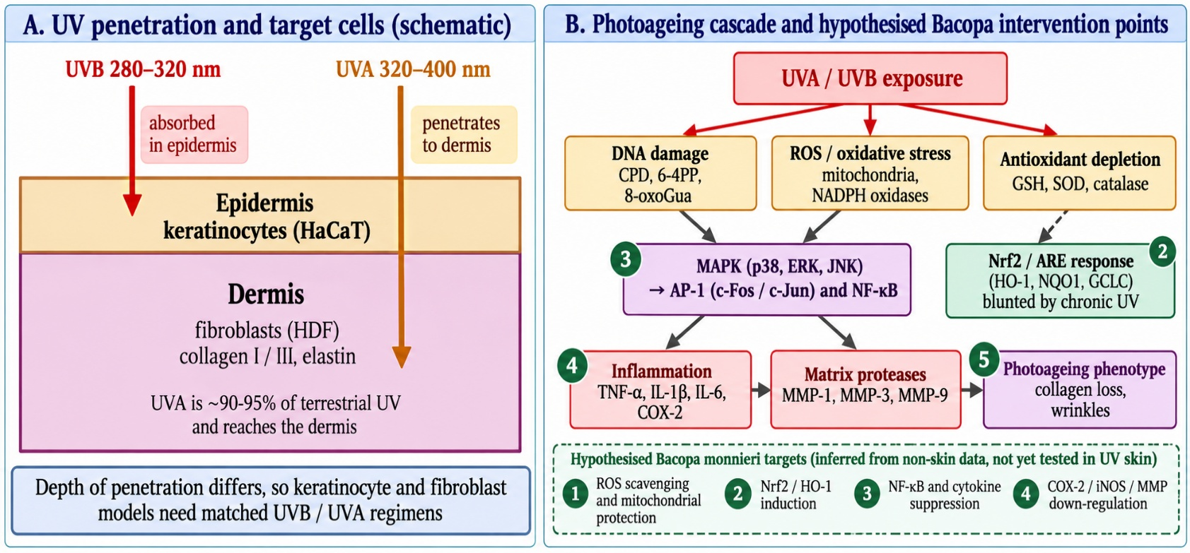
UVB (280–320 nm) is absorbed almost completely in the epidermis, whereas UVA (320–400 nm), which makes up the great majority of terrestrial UV, penetrates deeply into the dermis [8]. UVB forms cyclobutane pyrimidine dimers and 6-4 photoproducts directly in DNA; UVA is less energetic but roughly twenty times more intense in sunlight, and produces dimers together with oxidative lesions, chiefly 8-oxoguanine [9]. DNA damage is not merely a mutagenic event, since in human skin it also initiates release of matrix metalloproteinase-1 (MMP-1) [10]. The difference in penetration depth matters experimentally, because keratinocytes and dermal fibroblasts are exposed to different wavelengths in vivo (Figure 1, panel A).
2.2Oxidative stress and signalling
The skin generates reactive oxygen species (ROS) through mitochondrial respiration, NADPH oxidases, cyclooxygenase and lipoxygenase activity and Fenton chemistry, and UV is the largest contributor to extrinsic oxidative load [11]. UV simultaneously depletes cutaneous antioxidants, so that ROS accumulation is both increased and poorly buffered, and the resulting oxidative and inflammatory signals recruit neutrophil proteases that add to matrix damage [12]. In human skin in vivo, suberythemal UVB induced MMP mRNA, protein and activity within hours, while the transcription factors AP-1 and NF-κB were activated within minutes; pretreatment with retinoic acid blunted both responses [13]. The Nrf2/ARE axis, which drives expression of haem oxygenase-1, NAD(P)H quinone oxidoreductase and glutathione-synthesis enzymes, is the principal counterweight, and phytochemicals that activate it are being explored as photoprotectants [14].
2.3Inflammation, matrix degradation and repair
UV-induced MMPs degrade collagen and other matrix proteins, and in human skin all members of the MMP family respond to UV, with both epidermal and dermal contributions [15]. In reconstructed human skin, UVA triggered apoptosis of superficial dermal fibroblasts about six hours after exposure, with collagenase secretion and later repair of the dermis over two weeks, a sequence that links acute cell loss to the chronic phenotype [16]. Photoageing and chronological ageing share core molecular pathways, so that an agent that restrains UV-induced oxidative and inflammatory signalling may have wider anti-ageing relevance [1].
3Experimental models of UV-induced skin ageing
Three tiers of model are in routine use (Table 1). Monolayer HaCaT keratinocytes and primary or immortalised dermal fibroblasts permit rapid mechanistic work. In one such study, UVA at 10 J/cm² raised active MMP-1 in both cell types, and the test compound lowered it in a dose-dependent manner while suppressing p38, ERK and JNK phosphorylation, nuclear c-Fos and c-Jun, and TNF-α, COX-2, IL-1β and IL-6 [17]. In HaCaT cells irradiated with UVB at 30 mJ/cm², a plant fraction restored NRF2 and haem oxygenase-1, lowered ROS and dampened NF-κB activation [18]. Fibroblast models also capture senescence: UVA at 10 J/cm² increased senescence-associated β-galactosidase-positive cells to 23.07%, and pretreatment with 25 µg/mL EGCG reduced this to 12.75% together with lower MMPs and p66Shc [19].
Reconstructed skin adds tissue architecture and shows how UVA injures the dermal compartment while sparing keratinocytes [16]. In vivo, the outbred SKH1 hairless mouse is the most widely used strain because it is unpigmented and immunocompetent, although its poorly characterised genetic background and outbred status limit reproducibility [20]. Typical designs escalate UVB over weeks: oral Centella asiatica extract at 40 or 80 mg/kg/day was tested in Skh:HR-1 mice receiving UVB three times weekly, rising from 60 to 240 mJ/cm² over eight weeks, and reduced wrinkling, transepidermal water loss, malondialdehyde and cytokines while raising antioxidant enzymes and collagen expression, although several changes did not reach significance in post-hoc testing and some authors are affiliated with the extract's producer [21]. A topical design in hairless mice, daily UVB for 28 days, found that EGCG at 10 and 50 mg/mL, but not 1 mg/mL, reduced keratinocyte apoptosis and mitochondrial damage [22]. A polyherbal formula protected both human dermal fibroblasts (UVA 8 J/cm²) and BALB/c mice by raising nuclear Nrf2 and lowering 8-hydroxy-2'-deoxyguanosine [23].
These models answer different questions and should be chosen deliberately. Keratinocyte monolayers are suited to testing whether a candidate intercepts the earliest ROS, MAPK and NF-κB events after UVB, while fibroblast monolayers report on UVA-driven MMP expression, collagen turnover and senescence, which are the closest cellular correlates of wrinkling. Both lack the barrier, the vasculature and the cell-cell signalling of intact skin, and in vitro irradiation is often delivered through a thin layer of buffer after the medium has been removed, so that the effective dose and the timing of extract addition (before, during or after UV) shape the result. Rodent studies add pharmacokinetics, a measurable barrier and clinical-style endpoints, but the dose escalation schedule, the strain and the route of administration all vary between laboratories, and the hairless mouse differs from human skin in several respects, including its outbred genetic background [20]. Positive controls such as a commercial sunscreen [22] or a recognised antioxidant are therefore essential for putting any botanical effect in context.
| Model | UV regimen | Intervention | Reported endpoints | Ref. |
|---|---|---|---|---|
| HaCaT cells and human dermal fibroblasts | UVA 10 J/cm² | Syringaresinol 1, 5, 20 µM, 24 h | Active MMP-1 lowered at 20 µM (8.16 to 2.56 ng/mL in HaCaT; 2.28 to 1.37 ng/mL in fibroblasts); p38/ERK/JNK, c-Fos/c-Jun and cytokines reduced | [17] |
| Normal human dermal fibroblasts; BALB/c mice | UVA 8 J/cm² (cells); cumulative 60 J/cm² (mice) | Polyherbal extract 7.5–30 µg/mL; hesperetin | ROS and MMP-1 lower, collagen preserved; Nrf2, GST and NQO-1 higher; mouse nuclear Nrf2 up, 8-OHdG down | [23] |
| HaCaT keratinocytes | UVB 30 mJ/cm² | Plant fraction 100 µg/mL, 18 h | NRF2 and HO-1 restored; ROS reduced; NF-κB activation dampened | [18] |
| CCC-ESF-1 skin fibroblasts | UVA 10 J/cm² | EGCG 25 µg/mL, 24 h pretreatment | SA-β-gal-positive cells 23.07% to 12.75%; senescence markers, MMPs and p66Shc reduced | [19] |
| Skh:HR-1 hairless mice, 8 weeks | UVB 3 times weekly, 60 to 240 mJ/cm² | Oral Centella extract 40, 80 mg/kg/day | Wrinkles, TEWL, MDA and cytokines lower; antioxidant enzymes and collagen higher; MMP-1/-9 suppressed (not all significant) | [21] |
| Hairless mice, 28 days | UVB 100 µW/cm², 30 min daily | Topical EGCG 1, 10, 50 mg/mL; SPF 50 sunscreen control | Less keratinocyte apoptosis and mitochondrial damage at 10 and 50 mg/mL; little effect at 1 mg/mL | [22] |
| Reconstructed human skin | UVA | None (injury model) | Dermal fibroblast apoptosis at about 6 h, cells absent by 48 h; collagenase secretion; repair over 2 weeks | [16] |
| Any of the above | – | Bacopa monnieri extract or bacosides | No peer-reviewed study identified | – |
4Bacopa monnieri: Phytochemistry and extract variability
The bioactivity of Bacopa is usually attributed to its triterpenoid saponins, the bacosides, bacopasaponins and bacopasides, together with phenolic constituents and flavonoids. In a recently profiled ethanolic extract, bacopasaponin C reached 52.71 µg/mg and bacopaside II 43.05 µg/mg, and total phenolics were 9.27 mg gallic acid equivalents per gram [24]. Bioassay-guided work on ethyl acetate fractions isolated quercetin and isochlorogenic acid A [25], and quercetin was also the strongest binder to inflammatory targets among Bacopa constituents in a network-pharmacology study, in which it, rather than the whole extract, lowered nitric oxide and COX-2/iNOS expression in macrophages [26].
Extract composition therefore matters. A hexane extract protected HT-22 cells from glutamate toxicity and extended lifespan in Caenorhabditis elegans, whereas the ethanol extract did not [27]. Likewise a triterpenoid fraction and a bacoside-enriched fraction of the same methanolic extract differed, the latter showing stronger antioxidant activity and the former alone showing anti-arthritic effects [28]. Any skin study must therefore specify the solvent, the marker content and the standardisation, otherwise results cannot be compared between laboratories.
5Antioxidant evidence
The closest evidence to the UV context comes from an early study in which a methanol extract scavenged free radicals in a dose-dependent manner, protected DNA from cleavage produced by UV photolysis of hydrogen peroxide, and shielded non-immortalised human fibroblasts from peroxide-induced cytotoxicity and DNA damage [29]. This is a chemical and fibroblast system rather than irradiated skin, but it addresses two features of photoageing, radical generation and DNA injury, in a relevant cell type.
Mechanistic data come from neuronal models. In SK-N-SH cells exposed to hydrogen peroxide or acrolein, a standardised extract lowered intracellular ROS, preserved mitochondrial membrane potential and modulated NF-κB, Sirt1, ERK1/2 and p66Shc [30]. In PC12 cells challenged with sodium nitroprusside, the extract lowered nitric oxide and iNOS, restored antioxidant status, preserved mitochondrial membrane potential and reversed changes in Bax, Bcl-2, cytochrome c, caspase-3 and haem oxygenase-1 [31]. Mitochondrial protection and p66Shc modulation are noteworthy for skin because p66Shc is also lowered by EGCG in UVA-induced fibroblast senescence [19].
The Nrf2 pathway has been addressed in vivo. In rats given intracerebroventricular okadaic acid, a standardised extract at 40 and 80 mg/kg restored cortical and hippocampal Nrf2, haem oxygenase-1 and glutamate-cysteine ligase catalytic subunit expression and reduced oxidative stress, neuroinflammation and neuronal loss [32]. Direct in vitro radical-scavenging capacity of an extract was reported with IC50 values of 3.05 mg/mL (DPPH), 1.95 mg/mL (ABTS) and 0.59 mg/mL (superoxide) [24]. These potencies are modest, which suggests that cellular signalling effects, rather than stoichiometric scavenging, are more likely to explain any protective action. Finally, in skin-relevant tissue, a 50% ethanolic extract given orally at 25 mg/kg/day improved incision, excision and dead-space wound healing in rats, raised antioxidant levels and lowered free radicals and myeloperoxidase [33].
Comparators help to calibrate these findings. The okadaic acid study included melatonin at 20 mg/kg as a reference antioxidant [32], and the wound-healing study used an established antioxidant vitamin as its comparator [33], which at least places the extract within a recognisable efficacy range. No Bacopa study has yet used a sunscreen, a photoprotective retinoid or a validated botanical such as EGCG or Centella as a benchmark in an irradiated system, which is the comparison that a photoprotection claim would require [4,21].
6Anti-inflammatory evidence
Bacopa extracts act on innate immune cells at several levels. A bacoside-rich ethanolic extract lowered nitric oxide and TNF-α in stimulated RAW264.7 macrophages, with EC50 values of 0.25 and 1.15 mg/mL respectively, and lowered interferon-γ in stimulated human whole blood [34]. Triterpenoid and bacoside-enriched fractions each reduced TNF-α, IL-6 and nitrite in LPS-activated mononuclear cells and decreased carrageenan paw oedema in animals [28]. In activated N9 microglia, tea, infusion and alkaloid extracts and bacoside A inhibited TNF-α and IL-6 release, and in cell-free assays they inhibited caspase-1, caspase-3 and MMP-3 [35]. The last finding is directly pertinent to skin, since MMP-3 contributes to dermal matrix turnover; however, it is a cell-free observation and tells us nothing about expression in irradiated tissue.
Cytokine and enzyme effects are consistent across tissues. In TNF-α- and IFN-γ-primed SH-SY5Y cells an extract reduced IL-1β, IL-6, COX-2 and iNOS [36]. Bacoside A lowered IL-6, IL-17A, TNF-α and CCL-5 in mouse experimental autoimmune encephalomyelitis, but only the 10 mg/kg dose protected, and higher doses did not, a reminder that dose-response may be non-monotonic [37]. An oral extract at 50 mg/kg/day for 15 days lowered IL-6, TNF-α, MCP-1, COX-2, iNOS, ROS and nitric oxide in a colchicine model [38], and bacoside A lowered IL-1β, IL-6, TNF-α, NF-κB p65, COX-2 and iNOS activity in a 6-hydroxydopamine model of Parkinson's disease [39]. An ethyl acetate fraction inhibited leukotriene C4 synthase expression by 69% in HL-60 cells, inhibited LTA4 hydrolase and COX-2, and an oral extract suppressed ovalbumin-induced lung inflammation in mice [25]. In healthy older adults, three months of supplementation lowered NF-κB p65 phosphorylation [40], and a systematic review of twenty-two trials at 160–640 mg reported reduced NF-κB phosphorylation, inflammatory biomarkers and oxidative stress, although it addressed cognition, not skin [6].
Mechanistic interpretation should be cautious. The macrophage study proposed COX-2, NF-κB and the p38 and ERK kinases as likely upstream targets but did not test them [34], and several of the cytokine effects above were measured after a single stimulus in a single cell type. The pathways involved nevertheless coincide with those that UV engages in keratinocytes and fibroblasts, in which p38, ERK and JNK activation leads to c-Fos and c-Jun accumulation and to TNF-α, COX-2, IL-1β and IL-6 release [17]. It is this overlap, rather than any demonstrated skin effect, that justifies testing Bacopa in UV models. The antioxidant and anti-inflammatory evidence is summarized in Table 2.
| Model | Preparation / dose | Key finding | Relevance to photoaged skin | Ref. |
|---|---|---|---|---|
| DNA cleavage assay; human fibroblasts | Methanol extract | Radical scavenging; protection of DNA against H₂O₂ UV-photolysis and fibroblasts against H₂O₂ injury | Closest to UV-linked damage in a skin cell type | [29] |
| SK-N-SH cells | Standardised extract (pretreatment) | Lower ROS, preserved mitochondrial potential; NF-κB, Sirt1, ERK1/2 and p66Shc modulated | Redox signalling shared with UV response | [30] |
| Rat brain, okadaic acid | Standardised extract 40, 80 mg/kg | Nrf2, HO-1 and GCLC expression restored; less oxidative stress | Nrf2/ARE defence axis | [32] |
| RAW264.7 cells; human whole blood | Bacoside-rich ethanolic extract | NO and TNF-α lowered (EC50 0.25, 1.15 mg/mL); IFN-γ lowered | Innate cytokine response | [34] |
| LPS-activated mononuclear cells; rodent oedema | Triterpenoid and bacoside fractions | TNF-α, IL-6 and nitrite lowered; paw oedema reduced | Fraction-dependent activity | [28] |
| N9 microglia; cell-free assays | Tea, infusion, alkaloid extracts; bacoside A | TNF-α, IL-6 release lowered; caspase-1, caspase-3 and MMP-3 inhibited | MMP-3 relevant to dermal matrix | [35] |
| RAW264.7 cells; docking | Quercetin (Bacopa-derived) | Top binder to COX-2, iNOS, LOX, STAT3, CCR1, MMP9; NO and COX-2/iNOS lowered | COX-2 and MMP9 targets | [26] |
| HL-60 cells; OVA-sensitised mice | Ethyl acetate fraction | LTC4S expression inhibited by 69%; LTA4H and COX-2 inhibited | Eicosanoid pathway | [25] |
| Rat excision, incision and dead-space wounds | 50% ethanol extract, 25 mg/kg/day orally | Better wound healing; antioxidants raised; MPO and free radicals lowered | Skin repair with antioxidant effect (oral) | [33] |
| Human dermal fibroblasts | Methanol extract | Migration increased via α5β1 integrin and FAK phosphorylation | Fibroblast repair behaviour | [41] |
7Bridging the evidence to UV-induced skin ageing
Placed against the cascade in Figure 1 (panel B), the Bacopa data map onto four nodes. First, scavenging of radicals and protection of mitochondria and DNA in fibroblasts and neuronal cells [29–31] would act upstream, where UV-generated ROS are produced. Second, restoration of Nrf2, haem oxygenase-1 and related enzymes in vivo [32] addresses the defence that chronic UV erodes [14]. Third, lowered NF-κB signalling and cytokines [30,37,39,40] targets the transcriptional layer that Fisher and colleagues showed is activated within minutes of UVB [13]. Fourth, reduced COX-2, iNOS and MMP-3 activity [25,26,35] bears on the effector molecules that destroy collagen [15].
Skin-directed observations are sparse but consistent with this view. A methanol extract promoted human dermal fibroblast migration through α5β1 integrin and focal adhesion kinase, which relates to wound repair rather than inflammation [41], and oral extract improved wound healing with higher antioxidant levels in rats [33]. The Centella asiatica comparator is instructive, because a triterpenoid-rich extract produced an integrated antioxidant, anti-inflammatory and matrix-preserving profile in UVB-irradiated hairless mice [21], and a phospholipid-formulated Centella extract protected dermal fibroblasts from menadione-induced ROS and lipopolysaccharide-induced IL-6 and collagen loss, whereas the unformulated extract showed little activity in the same functional assays [42]. Bacopa is also a saponin-rich botanical, but similarity of class is not evidence of similar effect.
Several caveats limit translation. Most Bacopa studies used oral dosing and neural tissue, whose pharmacokinetics and cell biology differ from those of irradiated epidermis and dermis. Some reports have internal inconsistencies, for example a title referring to mice where the abstract describes rats [39], and the dose used in vitro (up to milligrams per millilitre for extracts) may exceed what can be achieved in the skin [24,34]. Efficacy was not uniform: in an LPS dementia model the extract lowered cortical IL-1β by only 6.65% and TNF-α by 17.37%, and did not significantly rescue behaviour [24].
A useful way to frame the next experiments is to state what would count against the hypothesis. If Bacopa acted mainly through radical scavenging, its protection should be lost when the extract is washed off before irradiation yet retained when present during exposure; if it acted through Nrf2, protection should require a pretreatment interval long enough for new protein synthesis and should disappear when Nrf2 is silenced, in keeping with the role of this pathway in botanical photoprotection [14,18]. If its effect were confined to suppressing cytokines, MMP-1 would fall without a change in ROS. These predictions are testable with the same endpoints used for the comparators in Table 1 and would distinguish a genuine photoprotective mechanism from non-specific cytotoxicity or from absorption of UV by the extract itself.
8Formulation considerations for topical use
Bacopa saponins and polyphenols are poorly suited to unmodified topical delivery, and delivery work is still at an early stage. The only skin-directed carrier identified is an ethosomal system of twelve ether-injection formulations with bacoside A as marker; the lead batch reported 89% entrapment, a 188 nm vesicle size and deeper dye penetration into goat skin than a hydroethanolic solution, although the paper shows internal inconsistencies between text and tables and makes unsupported claims about collagen and elastin, so its findings should be treated cautiously [43]. Other delivery systems explored for Bacopa, including liposomes, bilosomes, solid lipid nanoparticles and a phosphatidylcholine complex, were designed for neuroactive or oral use [7]. The phospholipid precedent for Centella [42] and the importance of extract composition [27] suggest that a standardised, phospholipid- or vesicle-based Bacopa preparation is the logical first topical candidate.
9Research gaps and proposed validation pathway
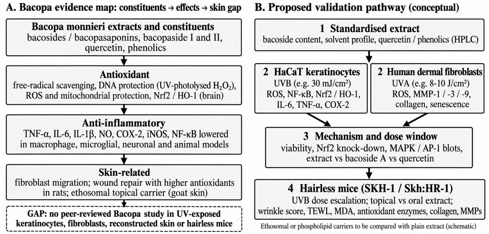
The central gap is the absence of any peer-reviewed study in UV-exposed keratinocytes, fibroblasts, reconstructed skin or hairless rodents. Within the literature we could reach there was also no Bacopa-specific Nrf2 study in skin cells and no clinical cosmetic study. A stepwise pathway is outlined in Figure 2 (panel B). A standardised extract, characterised by marker saponins and phenolic content, would first be tested in HaCaT cells under UVB and in dermal fibroblasts under UVA, with ROS, NF-κB, Nrf2/HO-1, MMP-1, -3 and -9 and collagen as readouts, using the UV doses and endpoints already established for comparators (Table 1) [17,18,23]. Mechanistic experiments should compare whole extract with bacoside A and quercetin and should include genetic or pharmacological Nrf2 inhibition. Promising preparations would then enter escalating-dose UVB protocols in hairless mice with topical and oral arms, wrinkle scoring, transepidermal water loss, malondialdehyde and antioxidant enzyme measurements [20,21]. Dose-response should be examined carefully, given the non-monotonic effect of bacoside A reported elsewhere [37].
Reporting quality will determine whether such studies are interpretable. Extract authentication, marker content, solvent, batch and vehicle should be stated, as the divergent activity of the hexane and ethanol extracts shows [27]. Experiments should include a vehicle control, an irradiated-only control, a non-irradiated extract control to exclude toxicity, and a reference photoprotector, and they should report significance for each endpoint rather than only the favourable ones, a point made necessary by the mixed statistical results in the Centella mouse study [21]. Because botanical extracts are often coloured and may absorb UV, optical interference with UV delivery and with colorimetric assays should be excluded.
10Conclusion
Bacopa monnieri has a credible mechanistic profile for photoprotection: it scavenges radicals, protects mitochondria, supports Nrf2 signalling and lowers NF-κB-dependent cytokines and COX-2/iNOS, and a cell-free assay shows inhibition of MMP-3. Every one of these observations, however, comes from non-cutaneous or non-irradiated systems. Until standardised extracts are tested in UVB-exposed keratinocytes, UVA-exposed fibroblasts and hairless mice against the endpoints established for other botanicals, Bacopa should be regarded as a promising but unproven candidate for UV-induced skin ageing.
Conflict of interest
The authors declare that there is no conflict of interest.
References
- [1]Fisher GJ, Kang S, Varani J, Bata-Csorgo Z, Wan Y, Datta S, et al. Mechanisms of photoaging and chronological skin aging. Arch Dermatol. 2002;138(11):1462–70. https://doi.org/10.1001/archderm.138.11.1462
- [2]Fisher GJ, Wang ZQ, Datta SC, Varani J, Kang S, Voorhees JJ. Pathophysiology of premature skin aging induced by ultraviolet light. N Engl J Med. 1997;337(20):1419–28. https://doi.org/10.1056/NEJM199711133372003
- [3]Krutmann J, Bouloc A, Sore G, Bernard BA, Passeron T. The skin aging exposome. J Dermatol Sci. 2017;85(3):152–61. https://doi.org/10.1016/j.jdermsci.2016.09.015
- [4]Nichols JA, Katiyar SK. Skin photoprotection by natural polyphenols: anti-inflammatory, antioxidant and DNA repair mechanisms. Arch Dermatol Res. 2010;302(2):71–83. https://doi.org/10.1007/s00403-009-1001-3
- [5]Zhao C, Wu S, Wang H. Medicinal plant extracts targeting UV-induced skin damage: molecular mechanisms and therapeutic potential. Int J Mol Sci. 2025;26(5):2278. https://doi.org/10.3390/ijms26052278
- [6]Valotto Neto LJ, Reverete de Araujo M, Moretti Junior RC, Mendes Machado N, Joshi RK, dos Santos Buglio D, et al. Investigating the neuroprotective and cognitive-enhancing effects of Bacopa monnieri: a systematic review focused on inflammation, oxidative stress, mitochondrial dysfunction, and apoptosis. Antioxidants (Basel). 2024;13(4):393. https://doi.org/10.3390/antiox13040393
- [7]Gościniak A, Stasiłowicz-Krzemień A, Szeląg M, Pawlak J, Skiera I, Kwiatkowska H, et al. Bacopa monnieri: preclinical and clinical evidence of neuroactive effects, safety of use and the search for improved bioavailability. Nutrients. 2025;17(11):1939. https://doi.org/10.3390/nu17111939
- [8]D'Orazio J, Jarrett S, Amaro-Ortiz A, Scott T. UV radiation and the skin. Int J Mol Sci. 2013;14(6):12222–48. https://doi.org/10.3390/ijms140612222
- [9]Cadet J, Douki T. Formation of UV-induced DNA damage contributing to skin cancer development. Photochem Photobiol Sci. 2018;17(12):1816–41. https://doi.org/10.1039/C7PP00395A
- [10]Dong KK, Damaghi N, Picart SD, Markova NG, Obayashi K, Okano Y, et al. UV-induced DNA damage initiates release of MMP-1 in human skin. Exp Dermatol. 2008;17(12):1037–44. https://doi.org/10.1111/j.1600-0625.2008.00747.x
- [11]Rinnerthaler M, Bischof J, Streubel MK, Trost A, Richter K. Oxidative stress in aging human skin. Biomolecules. 2015;5(2):545–89. https://doi.org/10.3390/biom5020545
- [12]Pillai S, Oresajo C, Hayward J. Ultraviolet radiation and skin aging: roles of reactive oxygen species, inflammation and protease activation, and strategies for prevention of inflammation-induced matrix degradation. Int J Cosmet Sci. 2005;27(1):17–34. https://doi.org/10.1111/j.1467-2494.2004.00241.x
- [13]Fisher GJ, Datta SC, Talwar HS, Wang ZQ, Varani J, Kang S, et al. Molecular basis of sun-induced premature skin ageing and retinoid antagonism. Nature. 1996;379(6563):335–9. https://doi.org/10.1038/379335a0
- [14]Chaiprasongsuk A, Panich U. Role of phytochemicals in skin photoprotection via regulation of Nrf2. Front Pharmacol. 2022;13:823881. https://doi.org/10.3389/fphar.2022.823881
- [15]Quan T, Qin Z, Xia W, Shao Y, Voorhees JJ, Fisher GJ. Matrix-degrading metalloproteinases in photoaging. J Investig Dermatol Symp Proc. 2009;14(1):20–4. https://doi.org/10.1038/jidsymp.2009.8
- [16]Bernerd F, Asselineau D. UVA exposure of human skin reconstructed in vitro induces apoptosis of dermal fibroblasts: subsequent connective tissue repair and implications in photoaging. Cell Death Differ. 1998;5(9):792–802. https://doi.org/10.1038/sj.cdd.4400413
- [17]Oh JH, Joo YH, Karadeniz F, Ko J, Kong CS. Syringaresinol inhibits UVA-induced MMP-1 expression by suppression of MAPK/AP-1 signaling in HaCaT keratinocytes and human dermal fibroblasts. Int J Mol Sci. 2020;21(11):3981. https://doi.org/10.3390/ijms21113981
- [18]Wang SH, Chen YS, Lai KH, Lu CK, Chang HS, Wu HC, et al. Prinsepiae Nux extract activates NRF2 activity and protects UVB-induced damage in keratinocyte. Antioxidants (Basel). 2022;11(9):1755. https://doi.org/10.3390/antiox11091755
- [19]Jia Y, Mao Q, Yang J, Du N, Zhu Y, Min W. (-)-Epigallocatechin-3-gallate protects human skin fibroblasts from ultraviolet A induced photoaging. Clin Cosmet Investig Dermatol. 2023;16:149–59. https://doi.org/10.2147/CCID.S398547
- [20]Benavides F, Oberyszyn TM, VanBuskirk AM, Reeve VE, Kusewitt DF. The hairless mouse in skin research. J Dermatol Sci. 2009;53(1):10–8. https://doi.org/10.1016/j.jdermsci.2008.08.012
- [21]Choi YJ, Cho EC, Lim S, Lee J, Bae J, Oh TK, et al. Oral Centella asiatica extract attenuates UVB-induced skin photoaging via antioxidant, anti-inflammatory, and extracellular matrix-preserving effects in hairless mice. Int J Mol Sci. 2026;27(1):204. https://doi.org/10.3390/ijms27010204
- [22]Liu C, Zheng X, Xiang L, Lu J, Polito CA, Liang Y. Protective effect of (-)-epigallocatechin gallate on ultraviolet B-induced skin damage in hairless mice. Trop J Pharm Res. 2016;15(6):1183–9. https://doi.org/10.4314/tjpr.v15i6.10
- [23]Lohakul J, Chaiprasongsuk A, Jeayeng S, Saelim M, Muanjumpon P, Thanachaiphiwat S, et al. The protective effect of polyherbal formulation, Harak Formula, on UVA-induced photoaging of human dermal fibroblasts and mouse skin via promoting Nrf2-regulated antioxidant defense. Front Pharmacol. 2021;12:649820. https://doi.org/10.3389/fphar.2021.649820
- [24]Olajide AT, Aunsorn S, Kehinde SA, Yazhen S, Kaewmanee T, Chusri S. Phytochemical profile, antioxidant activity, and neuroprotective effects of Bacopa monnieri extract in a lipopolysaccharide-induced dementia model. Int J Mol Sci. 2026;27(12):5229. https://doi.org/10.3390/ijms27125229
- [25]Soni KK, Shrivastava PN, Jones T, Mahady LJ, Patel U, Mukherjee P, et al. Extracts of Bacopa monnieri (L) Pennell down-regulate the expression of leukotriene C4 synthase mRNA in HL-60 cells and suppress OVA-induced inflammation in BALB/c mice. Curr Bioact Compd. 2014;10(1):21–30. https://doi.org/10.2174/157340721001140724152101
- [26]Jeyasri R, Muthuramalingam P, Adarshan S, Shin H, Ramesh M. Assessing the anti-inflammatory effects of Bacopa-derived bioactive compounds using network pharmacology and in vitro studies. ACS Omega. 2022;7(44):40344–54. https://doi.org/10.1021/acsomega.2c05318
- [27]Brimson JM, Prasanth MI, Plaingam W, Tencomnao T. Bacopa monnieri (L.) Wettst. extract protects against glutamate toxicity and increases the longevity of Caenorhabditis elegans. J Tradit Complement Med. 2020;10(5):460–70. https://doi.org/10.1016/j.jtcme.2019.10.001
- [28]Viji V, Helen A. Inhibition of pro-inflammatory mediators: role of Bacopa monniera (L.) Wettst. Inflammopharmacology. 2011;19(5):283–91. https://doi.org/10.1007/s10787-010-0046-4
- [29]Russo A, Izzo AA, Borrelli F, Renis M, Vanella A. Free radical scavenging capacity and protective effect of Bacopa monniera L. on DNA damage. Phytother Res. 2003;17(8):870–5. https://doi.org/10.1002/ptr.1061
- [30]Singh M, Murthy V, Ramassamy C. Modulation of hydrogen peroxide and acrolein-induced oxidative stress, mitochondrial dysfunctions and redox regulated pathways by the Bacopa monniera extract: potential implication in Alzheimer's disease. J Alzheimers Dis. 2010;21(1):229–47. https://doi.org/10.3233/JAD-2010-091729
- [31]Pandareesh MD, Anand T. Neuroprotective and anti-apoptotic propensity of Bacopa monniera extract against sodium nitroprusside induced activation of iNOS, heat shock proteins and apoptotic markers in PC12 cells. Neurochem Res. 2014;39(5):800–14. https://doi.org/10.1007/s11064-014-1273-7
- [32]Dwivedi S, Nagarajan R, Hanif K, Siddiqui HH, Nath C, Shukla R. Standardized extract of Bacopa monniera attenuates okadaic acid induced memory dysfunction in rats: effect on Nrf2 pathway. Evid Based Complement Alternat Med. 2013;2013:294501. https://doi.org/10.1155/2013/294501
- [33]Murthy S, Gautam MK, Goel S, Purohit V, Sharma H, Goel RK. Evaluation of in vivo wound healing activity of Bacopa monniera on different wound model in rats. Biomed Res Int. 2013;2013:972028. https://doi.org/10.1155/2013/972028
- [34]Williams R, Münch G, Gyengesi E, Bennett L. Bacopa monnieri (L.) exerts anti-inflammatory effects on cells of the innate immune system in vitro. Food Funct. 2014;5(3):517–20. https://doi.org/10.1039/c3fo60467e
- [35]Nemetchek MD, Stierle AA, Stierle DB, Lurie DI. The Ayurvedic plant Bacopa monnieri inhibits inflammatory pathways in the brain. J Ethnopharmacol. 2017;197:92–100. https://doi.org/10.1016/j.jep.2016.07.073
- [36]Costantini E, Jarlapoodi S, Serra F, Aielli L, Khan H, Belwal T, et al. Neuroprotective potential of Bacopa monnieri: modulation of inflammatory signals. CNS Neurol Disord Drug Targets. 2023;22(3):441–51. https://doi.org/10.2174/1871527321666220111124047
- [37]Madhu K, Prakash T, Maya S. Bacoside-A inhibits inflammatory cytokines and chemokine in experimental autoimmune encephalomyelitis. Biomed Pharmacother. 2019;109:1339–45. https://doi.org/10.1016/j.biopha.2018.10.188
- [38]Saini N, Singh D, Sandhir R. Bacopa monnieri prevents colchicine-induced dementia by anti-inflammatory action. Metab Brain Dis. 2019;34(2):505–18. https://doi.org/10.1007/s11011-018-0332-1
- [39]Zhang B, Shi J, Chang L, Wang H, Wang Y, Li M, et al. Bacoside-A exerts protective effect against Parkinson's disease-induced functional damage in mice via inhibition of apoptosis and oxidative response. Trop J Pharm Res. 2020;19(12):2565–70. https://doi.org/10.4314/tjpr.v19i12.12
- [40]Keegan AP, Stough C, Paris D, Luis CA, Abdullah L, Ait-ghezala G, et al. Bacopa monnieri supplementation has no effect on serum brain-derived neurotrophic factor levels but beneficially modulates nuclear factor kappa B and cyclic AMP response element-binding protein levels in healthy elderly subjects. J Clin Transl Res. 2023;9(1):50–8. https://doi.org/10.18053/jctres.09.202301.007
- [41]Zirmire RK, Saha D, Dey R, Tanimu H, Zaarour R, Bird D, et al. Bacopa monnieri phytochemicals regulate fibroblast cell migration via modulation of focal adhesions. iScience. 2024;27(4):109489. https://doi.org/10.1016/j.isci.2024.109489
- [42]Nezi P, Vella E, Riva A, Cicogni M, Cicaloni V, Salvini L, et al. In vitro antioxidant and collagen restoration activities of a standardized Centella asiatica L. extract formulated with phospholipids. Front Mol Biosci. 2026;13:1832045. https://doi.org/10.3389/fmolb.2026.1832045
- [43]Dwivedi D, Nimisha, Mishra N, Singh SP, Shukla AK. Performance evaluation of Bacopa monneri-loaded ethosomes for topical delivery. J Appl Nat Sci. 2024;16(3):1040–8. https://doi.org/10.31018/jans.v16i3.5435