Pharmacy
Comparative Hepatoprotective Evaluation of Calotropis gigantea Root and Portulaca oleracea Foliated-Stem Extracts, Alone and in Combination, Against Carbon Tetrachloride-Induced Hepatotoxicity in Wistar Rats
Department of Pharmacology, SHEAT College of Pharmacy, Varanasi, Uttar Pradesh, India (affiliated to Dr. A.P.J. Abdul Kalam Technical University, Lucknow, Uttar Pradesh, India)
Published October 2026
Abstract
Carbon tetrachloride (CCl4)-induced hepatotoxicity is a well-established experimental model for evaluating hepatoprotective agents, given its reproducible induction of oxidative hepatocellular injury via cytochrome P450-mediated bioactivation to the trichloromethyl free radical [3-6]. This study evaluated the comparative and combined hepatoprotective potential of methanolic extracts of Calotropis gigantea root and Portulaca oleracea foliated stem against CCl4-induced hepatotoxicity in Wistar rats. Extracts were obtained by cold maceration (6.30% w/w and 8.40% w/w yield respectively) and screened for phytoconstituents, both testing positive for flavonoids, alkaloids, glycosides, saponins and tannins. Acute oral toxicity testing (OECD 423) confirmed safety up to 2000 mg/kg, and doses of 100, 200 and 400 mg/kg were selected for the main study. Sixty male Wistar rats were divided into ten groups (n = 6): normal control, CCl4 toxic control, silymarin (50 mg/kg), three dose levels each of C. gigantea and P. oleracea, and a low-dose combination. CCl4 administration markedly elevated serum ALT, AST, ALP and bilirubin and hepatic malondialdehyde while depleting glutathione and body/liver-weight indices; both extracts dose-dependently attenuated these changes, with C. gigantea 400 mg/kg (74.6% hepatoprotection), P. oleracea 400 mg/kg (78.5%) and particularly the combination (85.4%, approaching silymarin's 87.7%) producing the most pronounced normalisation, corroborated by histopathological improvement from severe centrilobular necrosis to near-normal architecture. These findings support a synergistic, antioxidant-mediated hepatoprotective action for the combined extracts, comparable to standard silymarin therapy.
Keywords: Calotropis gigantea; Portulaca oleracea; hepatoprotective activity; carbon tetrachloride; oxidative stress; silymarin; polyherbal combination; Wistar rats
1Introduction
Liver disease accounts for close to two million deaths annually worldwide and remains among the leading causes of morbidity in both developed and developing regions, driven by viral hepatitis, alcohol use, metabolic dysfunction and exposure to hepatotoxic xenobiotics [1]. The liver's central role in xenobiotic biotransformation renders it uniquely vulnerable to chemically induced injury, and carbon tetrachloride (CCl4) remains the most extensively validated experimental hepatotoxicant for modelling this process [2,3]. Following absorption, CCl4 is bioactivated by hepatic cytochrome P450 2E1 to the trichloromethyl free radical (•CCl3), which combines with molecular oxygen to form the highly reactive trichloromethyl peroxy radical (•OOCCl3); both radicals initiate lipid peroxidation of hepatocyte membrane phospholipids, deplete endogenous antioxidant reserves and culminate in centrilobular necrosis, mirroring the oxidative pathophysiology of numerous clinically relevant hepatotoxic insults [3-6].
Despite the scale of this burden, pharmacotherapy for hepatocellular injury remains limited. Silymarin, a standardised flavonolignan complex from Silybum marianum, is the most widely used reference hepatoprotectant, acting through membrane stabilisation, free-radical scavenging and stimulation of hepatocyte regeneration, but its modest oral bioavailability and the absence of other approved agents sustain active interest in alternative, plant-derived hepatoprotective candidates [11-15].
Calotropis gigantea (L.) R. Br. (Apocynaceae/Asclepiadaceae), commonly known as giant milkweed or Madar, is a traditionally used medicinal shrub whose roots, latex and leaves have documented antioxidant, anti-inflammatory, analgesic and antimicrobial activity attributed to a rich cardenolide, flavonoid and triterpenoid content [16-20,24]. Portulaca oleracea L. (Portulacaceae), or purslane, is a widely distributed edible herb recognised for an unusually dense phytochemical profile — including flavonoids, phenolic alkaloids that are largely unique to the genus, omega-3 fatty acids and polysaccharides — underlying reported antioxidant, anti-inflammatory, analgesic, wound-healing and hypoglycaemic activities [25-32].
Although both species have been individually reported to possess antioxidant and hepatoprotective potential, no study has directly compared their root/stem extracts or examined whether their combination confers additive or synergistic protection — a rationale consistent with the broader polyherbal principle that phytoconstituents with complementary mechanisms (direct radical scavenging, metal chelation, enzyme induction) may together outperform single-agent therapy [10]. The present study therefore aimed to: (i) extract and phytochemically characterise C. gigantea root and P. oleracea foliated-stem extracts; (ii) establish their acute oral safety; and (iii) comparatively and combinatorially evaluate their hepatoprotective efficacy against CCl4-induced hepatotoxicity in Wistar rats using serum biochemical, hepatic oxidative-stress and histopathological endpoints, benchmarked against silymarin.
2Materials and methods
2.1Plant material, extraction and phytochemical screening
Fresh roots of Calotropis gigantea and foliated stems of Portulaca oleracea were collected, authenticated and shade-dried before mechanical pulverisation to coarse powder. Extracts were prepared by cold maceration: 500 g of each powdered material was macerated in methanol (1:5 w/v) for 48 h with intermittent agitation, repeated for two cycles, and the pooled filtrates concentrated under reduced pressure at 40-45°C using a rotary vacuum evaporator [33,34]. Percentage yield was calculated as (weight of dried extract / weight of powdered material) × 100. Each extract was subjected to standard qualitative phytochemical screening for alkaloids, flavonoids, glycosides, saponins, tannins/phenolics, carbohydrates, proteins/amino acids, triterpenoids/steroids and fixed oils using established colour and precipitation tests [33-35]. Analytical-grade CCl4, olive oil and methanol, a silymarin reference standard, and diagnostic kits for serum ALT, AST, ALP and bilirubin were procured from certified suppliers; all other chemicals and reagents used were of analytical grade.
2.2Experimental animals and ethical approval
Healthy adult male Wistar rats (200-250 g) were housed under standard laboratory conditions (12 h light/dark cycle, 22 ± 2°C, free access to standard pellet diet and water) and acclimatised for seven days prior to experimentation. All procedures were conducted in accordance with CPCSEA guidelines following Institutional Animal Ethics Committee (IAEC) approval [37].
2.3Acute oral toxicity study
Acute oral toxicity of both extracts was assessed per OECD Guideline 423 (acute toxic class method) using a limit dose of 2000 mg/kg body weight; animals were observed for mortality and behavioural/autonomic signs over 14 days. Both extracts were well tolerated with no mortality or overt toxicity at the limit dose, confirming a favourable safety margin and permitting selection of 100, 200 and 400 mg/kg as the low, medium and high doses for the main study [36].
2.4Experimental design and induction of hepatotoxicity
Sixty rats were randomly allocated to ten groups of six: Group I, normal control (vehicle only); Group II, toxic control (CCl4 only); Group III, standard (CCl4 + silymarin 50 mg/kg); Groups IV-VI, CCl4 + C. gigantea extract at 100, 200 and 400 mg/kg respectively; Groups VII-IX, CCl4 + P. oleracea extract at 100, 200 and 400 mg/kg respectively; and Group X, CCl4 + combination of C. gigantea (200 mg/kg) and P. oleracea (200 mg/kg). Hepatotoxicity was induced by a single intraperitoneal dose of CCl4 (1.5 mL/kg, 1:1 v/v in olive oil) on day 0; test extracts and silymarin, suspended in 1% w/v sodium carboxymethylcellulose and administered by oral gavage at a constant dosing volume of 10 mL/kg to minimise inter-group vehicle variability, were given once daily for 14 days beginning 24 h after CCl4 challenge.
2.5Biochemical and oxidative stress assays
On day 15, blood was collected and serum separated for estimation of alanine aminotransferase (ALT), aspartate aminotransferase (AST), alkaline phosphatase (ALP) and total bilirubin using standard diagnostic kits [38-41]. Percentage hepatoprotection was calculated from serum ALT as [(toxic control mean − treated mean) / (toxic control mean − normal control mean)] × 100. Body weight was recorded on days 0 and 15, and relative liver weight calculated as (liver weight / final body weight) × 100 following euthanasia. Liver tissue homogenates were used to estimate malondialdehyde (MDA) by the thiobarbituric acid reactive substances (TBARS) method [7] and reduced glutathione (GSH) by Ellman's DTNB method [8,9].
2.6Histopathological examination
Liver sections from representative animals of each group were fixed in 10% neutral buffered formalin, paraffin-embedded, sectioned at 5 μm and stained with haematoxylin and eosin for histopathological grading of necrosis, inflammatory infiltration and fatty change by a blinded observer, with severity graded semi-quantitatively (absent, mild, moderate, severe) relative to normal-control architecture.
2.7Statistical analysis
All data are expressed as mean ± SEM (n = 6) and were analysed by one-way ANOVA followed by Tukey's post-hoc multiple comparison test using standard statistical software, with p < 0.05 considered significant.
3Results and discussion
Results are presented sequentially from extraction and phytochemical characterisation through acute toxicity, biochemical and oxidative-stress evaluation to histopathological confirmation, in direct correspondence with the study's three stated aims. All data are reported as mean ± SEM (n = 6) unless otherwise specified, with statistical significance assessed at p < 0.05.
3.1Extraction yield and phytochemical screening
Cold maceration of 500 g powdered C. gigantea root yielded 31.5 g of dried methanolic extract (6.30% w/w), while 500 g of P. oleracea foliated stem yielded a markedly higher 42.0 g (8.40% w/w), consistent with the latter's higher soluble-solids and mucilage content (Table 1). Qualitative phytochemical screening confirmed both extracts to be rich in flavonoids, alkaloids, glycosides, saponins and tannins/phenolics — phytoconstituent classes with well-documented free-radical-scavenging and membrane-stabilising activity directly relevant to hepatoprotection [10,25-28]. Triterpenoids/steroids were detected only in C. gigantea, consistent with its established cardenolide/triterpenoid chemistry [16-20], while fixed oils and fatty acids were detected only in P. oleracea, consistent with the reported omega-3-rich lipid content of its foliated stems [27]. This complementary phytochemical profile — C. gigantea contributing triterpenoid/cardenolide-type constituents and P. oleracea contributing phenolic-alkaloid and fatty-acid constituents — provided a rational phytochemical basis for evaluating their combination.
| Parameter / Phytoconstituent | C. gigantea Root Extract | P. oleracea Foliated-Stem Extract |
|---|---|---|
| Percentage Yield (% w/w) | 6.30 | 8.40 |
| Flavonoids | + | + |
| Alkaloids | + | + |
| Glycosides | + | + |
| Saponins | + | + |
| Tannins & Phenolics | + | + |
| Carbohydrates | + | + |
| Proteins & Amino Acids | + | + |
| Triterpenoids / Steroids | + | − |
| Fixed Oils & Fatty Acids | − | + |
3.2Acute toxicity, body weight and relative liver weight
Both extracts were well tolerated at the OECD 423 limit dose of 2000 mg/kg with no mortality or overt behavioural toxicity, confirming a wide margin of safety and supporting selection of 100-400 mg/kg as pharmacologically relevant, sub-toxic doses for the hepatoprotection study [36]. CCl4 challenge produced the hallmark toxic phenotype of markedly reduced body-weight gain (8.2 ± 1.1 g versus 32.4 ± 1.8 g in normal controls) and substantially increased relative liver weight (4.6 ± 0.14% versus 3.1 ± 0.09%), reflecting hepatocellular oedema, inflammatory infiltration and impaired nutrient utilisation characteristic of acute CCl4 injury (Table 2) [3-6]. Co-administration of either extract dose-dependently attenuated both changes: body-weight gain rose from 14.6 ± 1.3 g (C. gigantea 100 mg/kg) to 23.7 ± 1.4 g (400 mg/kg), and from 16.1 ± 1.3 g to 24.9 ± 1.5 g across the corresponding P. oleracea doses, with relative liver weight normalising in parallel. The combination group (27.8 ± 1.5 g; 3.3 ± 0.10%) and silymarin (26.5 ± 1.5 g; 3.4 ± 0.10%) produced the most pronounced normalisation, approaching but not fully matching normal-control values, consistent with partial but substantial recovery of hepatic and systemic homeostasis within the 14-day treatment window.
| Group | Body Weight Gain (g) | Relative Liver Weight (%) | % Hepatoprotection (ALT-based) |
|---|---|---|---|
| Normal Control | 32.4 ± 1.8 | 3.1 ± 0.09 | — |
| Toxic Control (CCl4) | 8.2 ± 1.1 | 4.6 ± 0.14 | 0.0 |
| Silymarin 50 mg/kg | 26.5 ± 1.5 | 3.4 ± 0.10 | 87.7 |
| C. gigantea 100 mg/kg | 14.6 ± 1.3 | 4.2 ± 0.12 | 27.7 |
| C. gigantea 200 mg/kg | 19.8 ± 1.4 | 3.8 ± 0.11 | 53.8 |
| C. gigantea 400 mg/kg | 23.7 ± 1.4 | 3.5 ± 0.10 | 74.6 |
| P. oleracea 100 mg/kg | 16.1 ± 1.3 | 4.1 ± 0.12 | 33.8 |
| P. oleracea 200 mg/kg | 21.2 ± 1.4 | 3.7 ± 0.11 | 60.8 |
| P. oleracea 400 mg/kg | 24.9 ± 1.5 | 3.4 ± 0.10 | 78.5 |
| Combination (CG200+PO200) | 27.8 ± 1.5 | 3.3 ± 0.10 | 85.4 |
3.3Effect on serum biochemical parameters
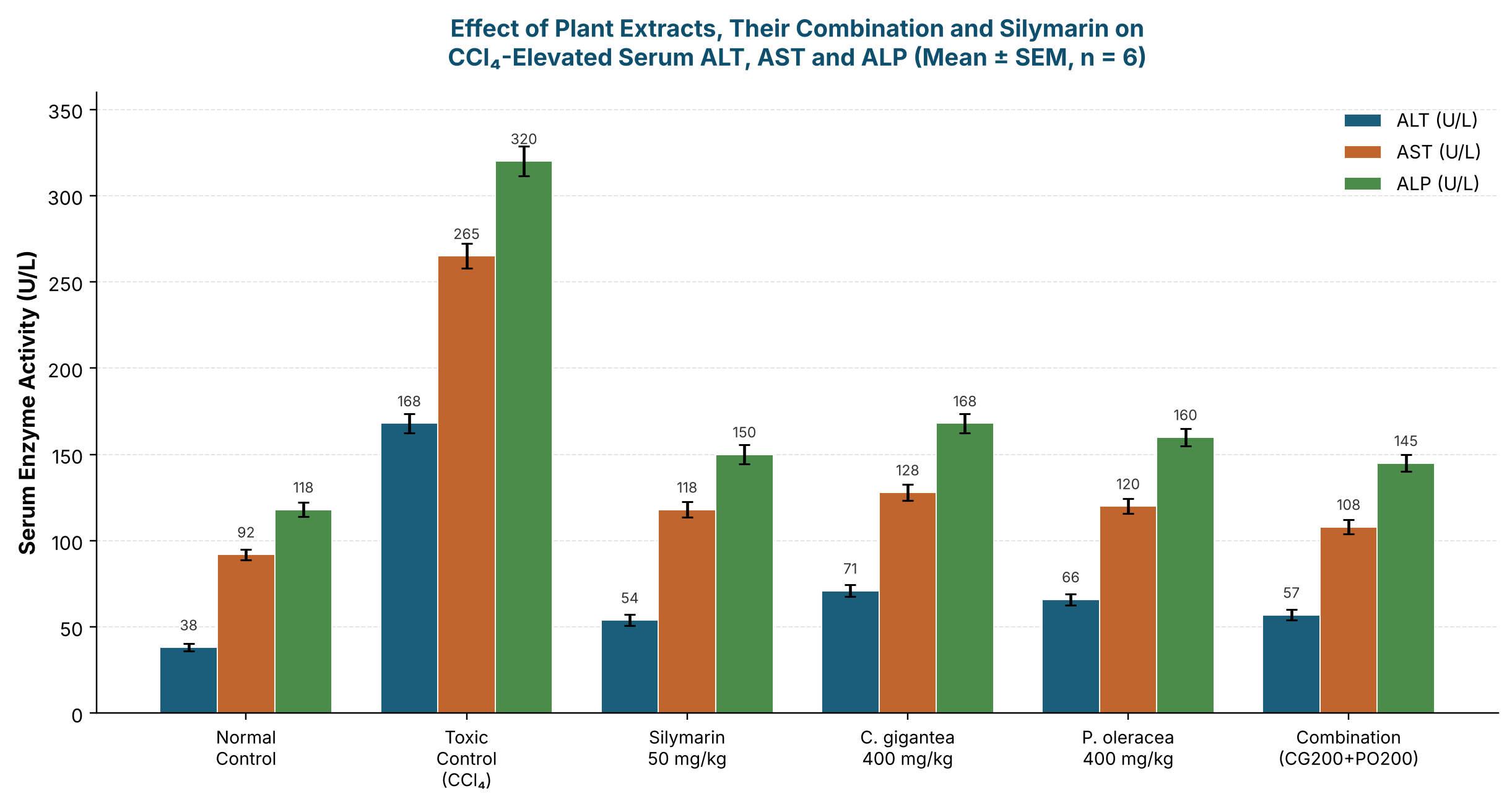
CCl4 administration produced marked hepatocellular leakage, elevating serum ALT from 38.2 ± 2.1 U/L (normal) to 168 ± 5.4 U/L, AST from 92 ± 3.0 to 265 ± 7.2 U/L, and ALP from 118 ± 4.2 to 320 ± 8.5 U/L (all p < 0.05 versus normal control), alongside a four-fold rise in total bilirubin (0.62 ± 0.04 to 2.45 ± 0.12 mg/dL), consistent with the expected CCl4 hepatotoxicity signature (Figure 1) [3,38-41]. Silymarin significantly reduced all four markers (ALT 54 ± 3.1; AST 118 ± 4.5; ALP 150 ± 5.5 U/L; bilirubin 0.85 ± 0.06 mg/dL; all p < 0.05 versus toxic control). Both plant extracts produced clear, dose-dependent reductions: C. gigantea lowered ALT from 132 ± 4.8 U/L at 100 mg/kg to 71 ± 3.5 U/L at 400 mg/kg, while P. oleracea lowered ALT from 124 ± 4.5 U/L to 66 ± 3.2 U/L across the same dose range, with parallel, statistically significant dose-dependent reductions in AST, ALP and bilirubin at every dose level relative to the toxic control (p < 0.05). At the highest dose tested, P. oleracea (400 mg/kg) numerically outperformed C. gigantea (400 mg/kg) on all four markers, while the low-dose combination (CG200+PO200) achieved the greatest reduction among treatment groups (ALT 57 ± 3.0; AST 108 ± 4.1; ALP 145 ± 5.0 U/L; bilirubin 0.80 ± 0.05 mg/dL), closely approaching silymarin and indicating an additive or synergistic benefit of combining the two extracts at half their individual high doses.
3.4Percentage hepatoprotection
Percentage hepatoprotection, calculated from ALT normalisation relative to the toxic control, rose monotonically with dose for both extracts (Table 2): from 27.7% to 74.6% for C. gigantea (100→400 mg/kg) and from 33.8% to 78.5% for P. oleracea across the same range, with P. oleracea consistently providing 2-6 percentage points greater protection than the equivalent C. gigantea dose. The combination achieved 85.4% hepatoprotection — exceeding either extract alone at its highest individual dose and approaching silymarin's 87.7% — despite each component being present at only half its top individual dose (200 mg/kg each versus 400 mg/kg alone), supporting a synergistic rather than purely additive interaction between the two extracts' complementary phytoconstituent profiles.
3.5Effect on hepatic oxidative stress markers
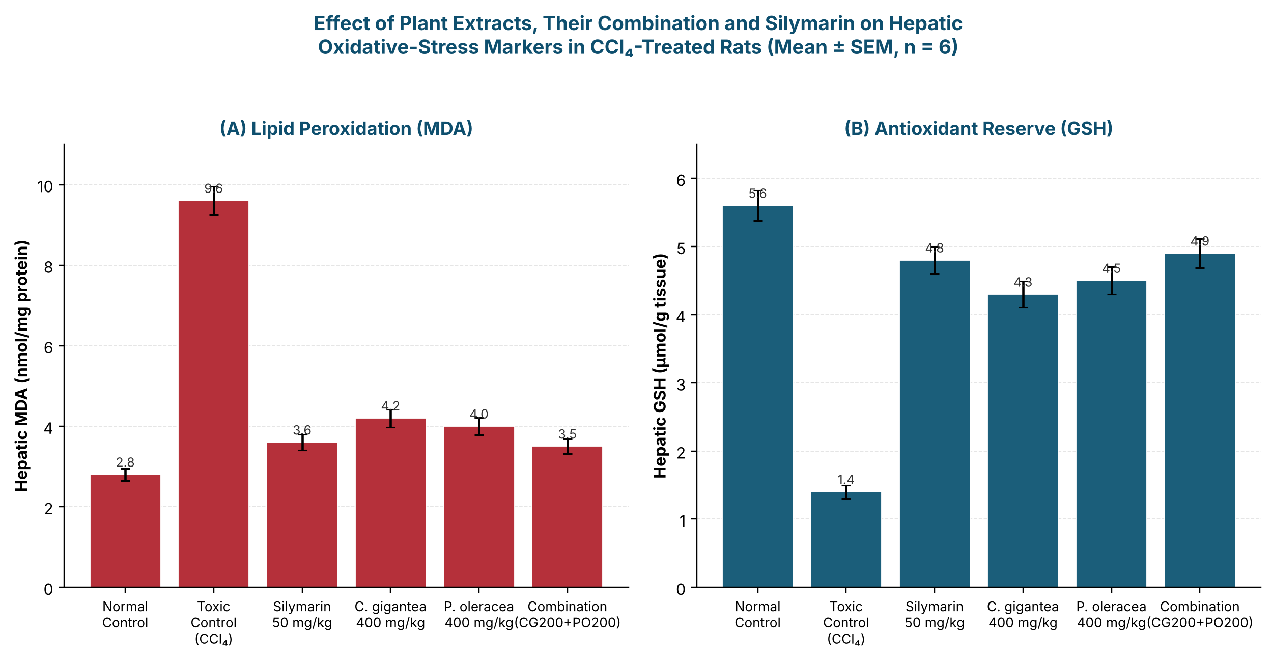
CCl4 intoxication produced the expected pro-oxidant hepatic phenotype: malondialdehyde (MDA), the principal end-product of lipid peroxidation, rose more than three-fold from 2.8 ± 0.15 to 9.6 ± 0.35 nmol/mg protein, while reduced glutathione (GSH), the primary non-enzymatic antioxidant buffer, fell by 75% from 5.6 ± 0.22 to 1.4 ± 0.10 μmol/g tissue (both p < 0.05 versus normal control), directly reflecting the free-radical-mediated membrane injury underlying the •CCl3/•OOCCl3 mechanism (Figure 2) [3-7,10]. Silymarin substantially restored both markers (MDA 3.6 ± 0.20; GSH 4.8 ± 0.20; p < 0.05 versus toxic control). Both extracts dose-dependently suppressed lipid peroxidation and restored glutathione reserve: C. gigantea reduced MDA from 7.5 ± 0.30 to 4.2 ± 0.22 nmol/mg protein (100→400 mg/kg) while raising GSH from 2.3 ± 0.14 to 4.3 ± 0.19 μmol/g, and P. oleracea produced a comparable, slightly stronger response (MDA 7.0 ± 0.28 to 4.0 ± 0.21; GSH 2.6 ± 0.15 to 4.5 ± 0.20) across the same dose range. The combination group showed the most favourable oxidative-stress profile among treatment groups (MDA 3.5 ± 0.19 nmol/mg protein; GSH 4.9 ± 0.21 μmol/g tissue), statistically indistinguishable from silymarin, reinforcing that the biochemical hepatoprotection observed in Figure 1 and Table 2 is mechanistically underpinned by restoration of the hepatic oxidant/antioxidant balance rather than a non-specific effect.
3.6Histopathological evaluation
Histopathological examination corroborated the biochemical findings (Table 3). Normal-control livers showed unremarkable architecture, whereas toxic-control sections displayed severe centrilobular necrosis, dense inflammatory infiltration and extensive fatty change, consistent with the marked enzyme elevations and oxidative-stress derangement reported above. Silymarin-treated livers showed only mild residual changes, confirming substantial histological recovery. Among the representative high-dose and combination groups selected for histopathological grading, both C. gigantea (400 mg/kg) and P. oleracea (400 mg/kg) markedly reduced necrosis, inflammation and fatty change to a mild grade, broadly comparable to silymarin, while the combination group showed the most complete recovery — minimal residual inflammation and fatty change with near-normal overall architecture — the best histological outcome among all extract-treated groups and consistent with its leading biochemical and oxidative-stress results.
| Group | Necrosis | Inflammation | Fatty Change | Overall Assessment |
|---|---|---|---|---|
| Normal Control | − | − | − | Normal |
| Toxic Control (CCl4) | +++ | +++ | +++ | Severe |
| Silymarin 50 mg/kg | + | ± | + | Mild |
| C. gigantea 400 mg/kg | + | + | + | Mild |
| P. oleracea 400 mg/kg | + | + | + | Mild |
| Combination (CG200+PO200) | ± | − | ± | Near-normal |
3.7Discussion
Collectively, these findings demonstrate that methanolic extracts of C. gigantea root and P. oleracea foliated stem each possess dose-dependent hepatoprotective activity against CCl4-induced hepatotoxicity across biochemical, oxidative-stress and histopathological endpoints, and that their combination — at half the individual high dose of each — matches or exceeds either extract alone and approaches standard silymarin therapy. This pattern is consistent with the complementary phytochemical profiles identified in Section 3.1: C. gigantea's triterpenoid/cardenolide-rich composition and P. oleracea's phenolic-alkaloid- and fatty-acid-rich composition may engage distinct but convergent protective mechanisms — direct radical scavenging, membrane stabilisation and modulation of cytochrome P450-mediated bioactivation — that together more effectively interrupt the CCl4 free-radical cascade than either mechanism alone [3-10,16-28]. The consistency between serum enzyme normalisation, restoration of hepatic MDA/GSH balance and histological recovery across all three endpoints strengthens confidence that the observed effects reflect genuine hepatoprotection rather than an isolated or artefactual biochemical change, and positions the C. gigantea-P. oleracea combination as a promising polyherbal candidate warranting further mechanistic and, ultimately, clinical investigation.
4Conclusion
This study demonstrates that methanolic extracts of Calotropis gigantea root and Portulaca oleracea foliated stem, both individually and in combination, confer significant, dose-dependent hepatoprotection against carbon tetrachloride-induced hepatotoxicity in Wistar rats, evidenced by normalisation of serum ALT, AST, ALP and bilirubin, restoration of hepatic MDA and GSH balance, and histopathological recovery from severe centrilobular necrosis toward near-normal architecture. P. oleracea was marginally more potent than C. gigantea at equivalent doses, but the combination of the two extracts at half their individual effective doses (200 mg/kg each) produced the greatest hepatoprotection among all tested regimens (85.4%), closely approaching silymarin (87.7%) and indicating a synergistic rather than purely additive interaction consistent with their complementary phytoconstituent profiles. These findings support further investigation of the C. gigantea-P. oleracea combination as a standardised polyherbal hepatoprotective formulation, including isolation and identification of the specific active constituents responsible for the observed synergy, and evaluation in chronic-dosing and dose-optimisation models prior to any translational development.
References
- [1]Asrani SK, Devarbhavi H, Eaton J, Kamath PS. Burden of liver diseases in the world. J Hepatol. 2019;70(1):151–171.
- [2]Bishayee A, Sarkar A, Chatterjee M. Hepatoprotective activity of carrot (Daucus carota L.) against carbon tetrachloride intoxication in mouse liver. J Ethnopharmacol. 1995;47(2):69–74.
- [3]Weber LW, Boll M, Stampfl A. Hepatotoxicity and mechanism of action of haloalkanes: carbon tetrachloride as a toxicological model. Crit Rev Toxicol. 2003;33(2):105–136.
- [4]Recknagel RO, Glende EA, Dolak JA, Waller RL. Mechanisms of carbon tetrachloride toxicity. Pharmacol Ther. 1989;43(1):139–154.
- [5]Manibusan MK, Odin M, Eastmond DA. Postulated carbon tetrachloride mode of action: a review. J Environ Sci Health C. 2007;25(3):185–209.
- [6]Slater TF. Free-radical mechanisms in tissue injury. Biochem J. 1984;222(1):1–15.
- [7]Ohkawa H, Ohishi N, Yagi K. Assay for lipid peroxides in animal tissues by thiobarbituric acid reaction. Anal Biochem. 1979;95(2):351–358.
- [8]Ellman GL. Tissue sulfhydryl groups. Arch Biochem Biophys. 1959;82(1):70–77.
- [9]Sedlak J, Lindsay RH. Estimation of total, protein-bound, and nonprotein sulfhydryl groups in tissue with Ellman's reagent. Anal Biochem. 1968;25:192–205.
- [10]Halliwell B, Gutteridge JM. Free Radicals in Biology and Medicine. 5th ed. Oxford: Oxford University Press; 2015.
- [11]Girish C, Koner BC, Jayanthi S, Rao KR, Rajesh B, Pradhan SC. Hepatoprotective activity of six polyherbal formulations in CCl4-induced liver toxicity in mice. Indian J Exp Biol. 2009;47(4):257–263.
- [12]Pradhan SC, Girish C. Hepatoprotective herbal drug, silymarin from experimental pharmacology to clinical medicine. Indian J Med Res. 2006;124(5):491–504.
- [13]Saller R, Meier R, Brignoli R. The use of silymarin in the treatment of liver diseases. Drugs. 2001;61(14):2035–2063.
- [14]Abenavoli L, Capasso R, Milic N, Capasso F. Milk thistle in liver diseases: past, present, future. Phytother Res. 2010;24(10):1423–1432.
- [15]Surai PF. Silymarin as a natural antioxidant: an overview of the current evidence and perspectives. Antioxidants (Basel). 2015;4(1):204–247.
- [16]Ramachandra Setty S, Quereshi AA, Viswanath Swamy AH, et al. Hepatoprotective activity of Calotropis procera flowers against paracetamol-induced hepatic injury in rats. Fitoterapia. 2007;78(7–8):451–454.
- [17]Kumar G, Karthik L, Rao KV. A review on pharmacological and phytochemical properties of Calotropis procera (Ait.) R. Br. (Asclepiadaceae). Asian J Pharm Clin Res. 2011;4(2):1–7.
- [18]Basu A, Chaudhuri AK. Preliminary studies on the antiinflammatory and analgesic activities of Calotropis procera root extract. J Ethnopharmacol. 1991;31(3):319–324.
- [19]Sharma AK, Kharb R, Kaur R. Pharmacognostical aspects of Calotropis gigantea (L.) R. Br. Int J Pharm Pharm Sci. 2011;3(2):15–17.
- [20]Rathod NR, Raghuveer I, Chitme HR, Chandra R. Free radical scavenging activity of Calotropis gigantea root bark. Pharmacologyonline. 2008;1:378–387.
- [21]Kumar S, Dewan S, Sangraula H, Kumar VL. Antidiarrhoeal activity of the latex of Calotropis procera. J Ethnopharmacol. 2001;76(1):115–118.
- [22]Dewan S, Sangraula H, Kumar VL. Preliminary studies on the analgesic activity of latex of Calotropis procera. J Ethnopharmacol. 2000;73(1–2):307–311.
- [23]Nenaah G. Antibacterial and antifungal activities of (E,E)-α-farnesene isolated from the aerial parts of Calotropis procera. J Taibah Univ Sci. 2013;7(4):197–204.
- [24]Rastogi S, Kumar R, Chaudhary GN, Mishra V. Phytochemical screening and GC-MS profiling of Calotropis gigantea extracts. J Pharmacogn Phytochem. 2016;5(3):200–205.
- [25]Xiang L, Xing D, Wang W, Wang R, Ding Y, Du L. Alkaloids from Portulaca oleracea L. Phytochemistry. 2005;66(21):2595–2601.
- [26]Yang Z, Liu C, Xiang L, Zheng Y. Phenolic alkaloids as a new class of antioxidants in Portulaca oleracea. Phytother Res. 2009;23(7):1032–1035.
- [27]Uddin MK, Juraimi AS, Hossain MS, Un A, Ali M, Rahman MM. Purslane weed (Portulaca oleracea): a prospective plant source of nutrition, omega-3 fatty acid, and antioxidant attributes. ScientificWorldJournal. 2014;2014:951019.
- [28]Zhu H, Wang Y, Liu Y, Xia Y, Tang T. Analysis of flavonoids in Portulaca oleracea L. by UV-vis spectrophotometry with comparative study on different extraction technologies. Food Anal Methods. 2010;3(2):90–97.
- [29]Chan K, Islam MW, Kamil M, et al. The analgesic and anti-inflammatory effects of Portulaca oleracea L. subsp. sativa (Haw.) Celak. J Ethnopharmacol. 2000;73(3):445–451.
- [30]Gong F, Li F, Zhang L, Li J, Zhang Z, Wang G. Hypoglycemic effects of crude polysaccharide from Purslane (Portulaca oleracea L.) in normal and diabetic mice. Molecules. 2009;14(12):4844–4854.
- [31]Rashed AN, Afifi FU, Disi AM. Simple evaluation of the wound healing activity of a crude extract of Portulaca oleracea L. (growing in Jordan) in Mus musculus JVI-1. J Ethnopharmacol. 2003;88(2–3):131–136.
- [32]Erkan N. Antioxidant activity and phenolic compounds of fractions from Portulaca oleracea L. Food Chem. 2012;133(3):775–781.
- [33]Harborne JB. Phytochemical Methods: A Guide to Modern Techniques of Plant Analysis. 3rd ed. London: Chapman and Hall; 1998.
- [34]Evans WC. Trease and Evans' Pharmacognosy. 16th ed. Edinburgh: Saunders Elsevier; 2009.
- [35]Sofowora A, Ogunbodede E, Onayade A. The role and place of medicinal plants in the strategies for disease prevention. Afr J Tradit Complement Altern Med. 2013;10(5):210–229.
- [36]Organisation for Economic Co-operation and Development. Test No. 423: Acute Oral Toxicity - Acute Toxic Class Method. OECD Guidelines for the Testing of Chemicals, Section 4. Paris: OECD Publishing; 2002.
- [37]Committee for the Purpose of Control and Supervision of Experiments on Animals (CPCSEA). CPCSEA Guidelines for Laboratory Animal Facility. Indian J Pharmacol. 2003;35:257–274.
- [38]Ozer J, Ratner M, Shaw M, Bailey W, Schomaker S. The current state of serum biomarkers of hepatotoxicity. Toxicology. 2008;245(3):194–205.
- [39]Giannini EG, Testa R, Savarino V. Liver enzyme alteration: a guide for clinicians. CMAJ. 2005;172(3):367–379.
- [40]Amacher DE. Serum transaminase elevations as indicators of hepatic injury following the administration of drugs. Regul Toxicol Pharmacol. 1998;27(2):119–130.
- [41]Ahmed B, Alam T, Varshney M, Khan SA. Hepatoprotective activity of two plants belonging to the Apiaceae and the Euphorbiaceae family. J Ethnopharmacol. 2002;79(3):313–316.