Pharmacy
Chitosan-Coated Quercetin Lipid Nanoparticles for Intranasal Brain Delivery: A Short Review
1Department of Pharmaceutics, S. N. College of Pharmacy, Babupur, Lucknow Road, Jaunpur, Uttar Pradesh, India
2Department of Pharmaceutics, S. N. College of Pharmacy, Babupur, Lucknow Road, Jaunpur, Uttar Pradesh, India (affiliated to Dr. A. P. J. Abdul Kalam Technical University, Lucknow, India)
Published October 2026
Abstract
Quercetin is a dietary flavonol with well-documented antioxidant and anti-inflammatory activity, and recent studies show that it can also lower amyloid burden, oxidative stress and neuroinflammation in animal models of Alzheimer's disease, Parkinson's disease and stress-related disorders. Its clinical use is limited by very low aqueous solubility (about 0.01 mg/mL), extensive intestinal and hepatic conjugation, and poor access to the brain across the blood–brain barrier. The intranasal route can deliver drugs to the brain along the olfactory and trigeminal nerves and bypasses hepatic first-pass metabolism, but mucociliary clearance removes most conventional nasal formulations within about 10 to 20 minutes. Lipid nanoparticles, particularly nanostructured lipid carriers (NLCs), solubilise lipophilic drugs and protect them from degradation, and coating them with the cationic polysaccharide chitosan adds mucoadhesion and a transient opening of tight junctions. Dispersing the coated particles in a thermosensitive in situ gel can extend residence time further. This short review draws on open online literature to describe the biopharmaceutical problem, the nose-to-brain pathways, lipid nanoparticle design, the intranasal quercetin nanocarriers reported so far, the rationale for chitosan functionalisation, in situ gelling vehicles, and a quality-by-design strategy using a three-factor Box–Behnken design. A staged evaluation plan covering physicochemical, in vitro, ex vivo, safety, stability and in vivo studies is proposed. No study identified in this review combined statistical optimisation, chitosan surface functionalisation of a lipid carrier, quantitative mucoadhesion and ciliotoxicity testing, and an in situ gel for quercetin in one integrated programme, and this gap is the main opportunity identified.
Keywords: quercetin; nanostructured lipid carriers; intranasal delivery; nose-to-brain; chitosan; mucoadhesion; in situ gel; Box-Behnken design; quality by design
1Introduction
Alzheimer's disease, Parkinson's disease and related neurodegenerative conditions share several features, notably oxidative stress, neuroinflammation and abnormal protein aggregation. For this reason plant-derived antioxidants and anti-inflammatory compounds have attracted interest as preventive or adjunct measures [1,2]. Quercetin (3,3′,4′,5,7-pentahydroxyflavone) is among the most widely studied of these compounds. It is present in onions, apples, berries and tea, and it scavenges free radicals, chelates metal ions and modulates inflammatory signalling [1,2]. Recent intranasal studies in rodents report that quercetin-loaded carriers improved cognition, restored antioxidant enzymes and reduced markers of neuroinflammation [3–5].
The difficulty is delivery, not pharmacology. Quercetin dissolves in water only to about 0.01 mg/mL at 25 °C, is metabolised extensively by glucuronidation, sulfation and methylation in the gut wall, and is chemically unstable at basic pH and under ultraviolet light [6]. Reviews of human studies describe low oral bioavailability that varies widely between individuals [7]. Even the fraction that reaches the circulation must then cross the blood–brain barrier, which excludes most large or poorly permeable molecules from the brain.
Intranasal administration offers a non-invasive way around both barriers. The nasal mucosa is richly vascularised, so absorbed drug avoids first-pass hepatic metabolism, and the olfactory and trigeminal nerves provide direct connections between the nasal cavity and the central nervous system [8,9]. A recent review of the field reports that intranasal dosing has produced brain exposure many times higher than oral dosing in comparative studies, and that dihydroergotamine and sumatriptan are already used intranasally for migraine with specialised devices [9]. The obstacle is the short residence time of a liquid in the nasal cavity, where mucociliary clearance operates on a timescale of roughly 10 to 20 minutes [10,11].
Nanostructured lipid carriers, chitosan coating and in situ gelling vehicles each address part of this problem. Lipid nanoparticles improve solubilisation and stability; chitosan sticks to negatively charged mucus and loosens epithelial tight junctions; and a gel that forms at body temperature reduces drainage [10,12,13]. Whether these three features can be combined and optimised systematically for quercetin is the question that motivates this review.
The article was compiled from open online resources, including publisher platforms, review articles and regulatory guidelines. Section 2 describes quercetin and its limitations. Section 3 covers the nose-to-brain route. Sections 4 and 5 deal with lipid nanoparticles and with the intranasal quercetin carriers reported so far. Sections 6 and 7 discuss chitosan functionalisation and in situ gels. Sections 8 and 9 describe optimisation and evaluation, and Sections 10 and 11 discuss challenges and conclusions.
2Quercetin: Pharmacology and biopharmaceutical limitations
2.1Neuroprotective pharmacology
Reviews of quercetin describe antioxidant, anti-inflammatory, antiviral, anticancer and cardioprotective activities, together with pharmacokinetic limitations that constrain clinical use [1,2]. In the nervous system, the effects most relevant to neurodegeneration are the reduction of oxidative damage, the suppression of pro-inflammatory cytokines and the interference with amyloid aggregation. Experimental support comes from the intranasal Alzheimer's model of Satpathy and colleagues, in which quercetin-loaded NLCs raised superoxide dismutase, glutathione and total antioxidant capacity, lowered malondialdehyde and acetylcholinesterase activity, reduced amyloid-β accumulation and improved spatial learning in streptozotocin-treated rats [3]. Transferrin-functionalised quercetin lipid nanoparticles inhibited fibril formation in vitro [14], and intranasal quercetin–chitosan nanoparticles restored hippocampal synaptic proteins and lowered inflammatory markers in a mouse model of post-traumatic stress disorder [5].
2.2Physicochemical and biopharmaceutical profile
Quercetin is a lipophilic flavonol with five hydroxyl groups. Its water solubility is about 0.01 mg/mL at 25 °C [6]. It is more stable in mildly acidic medium (around pH 5) than at pH 8, and exposure to UVA and UVB radiation produces degradation products [6]. These properties matter for formulation: a nasal product must be protected from light, and the acidic conditions used to dissolve chitosan for coating are not unfavourable for the drug. After oral absorption, about 90 % of the quercetin taken up by enterocytes is reported to be conjugated in the gut, so little free aglycone reaches the blood [6]. The human literature also stresses large differences in bioavailability between individuals [7].
Because quercetin is lipophilic and poorly water-soluble, it is expected to partition into the lipid phase of an NLC rather than the aqueous phase, and solubility screening in candidate solid and liquid lipids is therefore the first quantitative step in formulation development. Analytical methods must also be validated before any of the later measurements can be trusted, and the ICH Q2(R1) guideline sets out the parameters to be addressed, including linearity, accuracy, precision, specificity, detection and quantitation limits and robustness [15].
2.3The case for nanocarriers
A review of quercetin nanotechnology identifies seven classes of carriers, including liposomes, lipid nanoparticles, polymeric nanoparticles, cyclodextrins and biopolymeric systems based on chitosan, and notes that lipid nanoparticles improve gastrointestinal absorption and brain permeability, particularly when the surface is functionalised [6]. For a drug that must reach the brain, the more direct question is whether the carrier can be delivered by a route that avoids the gut and the liver. This leads to the nasal route.
3The nose-to-brain route: Pathways and barriers
3.1Anatomy and transport pathways
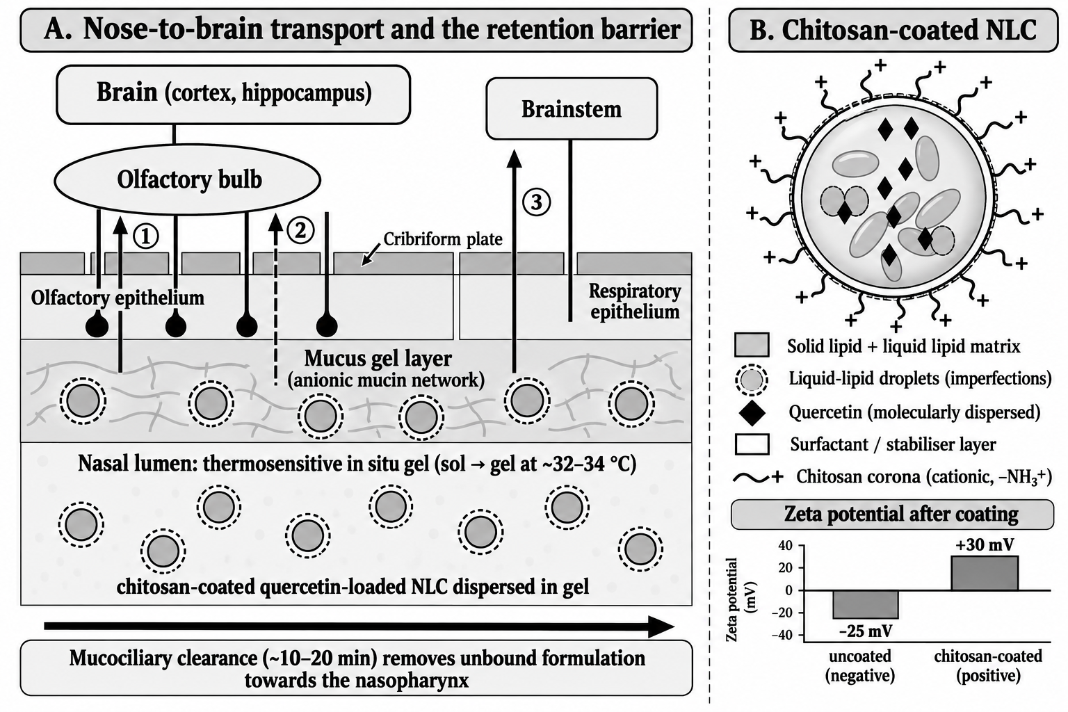
The nasal cavity contains a vestibule, an atrium, a respiratory region and an olfactory region. The olfactory epithelium contains sensory neurons whose axons pass through the cribriform plate to the olfactory bulb, while branches of the trigeminal nerve innervate the respiratory mucosa and project to the brainstem [9,10]. Together they form pathways that bypass the blood–brain barrier. A recent review describes two olfactory mechanisms: an intracellular route in which substances are taken up by olfactory neurons and carried along the axon, and an extracellular route along perineural and perivascular spaces, in which cerebrospinal fluid concentrations can peak within minutes [9]. The trigeminal route is described as slower and mainly transcellular [9]. A third possibility is absorption into the systemic circulation followed by limited transfer across the blood–brain barrier. These routes are shown in Figure 1 (panel A).
Which route dominates depends on the carrier. The olfactory pathway is described as suitable for particles smaller than about 200 nm [10], and particle size has a measurable effect on brain access: phenytoin-loaded NLCs below 50 nm gave higher cerebrospinal fluid concentrations than particles above 100 nm [10]. Higher lipophilicity and lower molecular weight of the drug are also associated with higher cerebrospinal fluid levels [9]. Formulation pH should lie between about 4.5 and 6.5 to protect the mucosa and ciliary function [9].
3.2Mucus, mucociliary clearance and the residence-time problem
The nasal epithelium is covered by a mucus gel that is moved towards the nasopharynx by ciliary beating. Reported clearance times are about 10 to 20 minutes, and the human mean transport rate has been quoted as about 6 mm per minute [9,10]. A detailed review of the olfactory mucosa treats mucociliary clearance and the small olfactory surface area as the principal formulation constraints for nose-to-brain transfer [17]. Mucin is an anionic glycoprotein network, and Illum's classic analysis identified rapid clearance as the main problem for nasal products and bioadhesive and gel-forming systems as the main solutions [11].
Formulation therefore matters as much as the drug. In studies summarised by one review, gels gave higher brain concentrations than solutions, thermoresponsive gels sustained those concentrations, and nanoparticles increased brain bioavailability about five-fold compared with free drug [9]. Lipid-based nanoparticles have shown drug-targeting efficiencies of several hundred per cent in some cases: donepezil-loaded SLNs gave a drug-targeting efficiency of 553.95 % and a direct-transport percentage of 81.94 %, and huperzine A nanoemulsions modified with lactoferrin gave a targeting efficiency of 320 % [10]. These numbers are not directly comparable across drugs, but they explain why lipid carriers are the most frequently studied class of nose-to-brain nanocarrier.
The size of the potential gain is illustrated by comparative pharmacokinetic data collected in one review, which report brain exposure after intranasal dosing that was 8 to 12 times higher than after subcutaneous dosing, about 25-fold higher than after oral dosing and 14.7 times higher than after intravenous dosing for the compounds analysed [9]. These are pooled literature values for different drugs and should not be applied to quercetin, but they show why the nasal route is being pursued for compounds that cannot cross the blood–brain barrier efficiently.
4Lipid nanoparticles: Structure, composition and manufacture
4.1From SLN to NLC
Solid lipid nanoparticles are made entirely of solid lipids. Their ordered crystalline matrix gives physical stability but can expel the drug during storage as the lipid recrystallises, and it limits the loading capacity [12,18]. Nanostructured lipid carriers solve this problem by blending a solid lipid with a spatially incompatible liquid lipid. The mixture produces a deliberately imperfect matrix with more space for drug molecules, higher payload, and reduced expulsion on storage [12,18]. This is the rationale for choosing an NLC rather than an SLN for a lipophilic flavonoid such as quercetin.
In the quercetin literature both types have been studied. Solid lipid nanoparticles of quercetin made from Compritol with Tween 80 and optimised with a central composite design were below 200 nm, entrapped 85.73 % of the drug and improved memory retention in rats compared with free quercetin [19]. In a later study, quercetin was encapsulated in transferrin-functionalised SLNs and NLCs of about 200 nm with entrapment of 80 to 90 %, and the NLC version showed better permeability across a blood–brain barrier model [14].
Lipid nanoparticles have further advantages for a molecule such as quercetin. They are made from lipids that are physiologically tolerated, they can protect a labile active from chemical and enzymatic degradation, and they can be dried or otherwise processed for long-term storage [18]. Localisation of quercetin within a lipid matrix would be expected to shield the flavonoid from light and from oxidation, a benefit given its known photodegradation [6], although this protective effect has to be demonstrated experimentally for each formulation.
4.2Composition
A typical NLC consists of a solid lipid, a liquid lipid and one or more surfactants. Candidate materials for a quercetin NLC include glyceryl behenate or glyceryl palmitostearate as solid lipids, oleic acid or Capryol 90 as liquid lipids, and poloxamer 188 or polysorbate 80 with soya lecithin as stabilisers. Published intranasal NLCs use comparable combinations. For example, a lurasidone NLC for intranasal use contained glyceryl monostearate (3.2 %), Capryol 90 (4.4 %) and Tween 80 (0.4 %) [20], and the intranasal quercetin NLC of Satpathy and colleagues used lecithin, sunflower oil and Cremophor RH 80 [3]. Final choices should rest on solubility of quercetin in each lipid and on compatibility studies, since the lipid phase dissolves the drug and controls release [18].
4.3Manufacture by hot homogenisation and ultrasonication
The hot homogenisation approach melts the lipids above the melting point of the solid lipid, dissolves or disperses the drug in the melt, and disperses that phase in a hot aqueous surfactant solution with high-shear mixing to form a coarse emulsion. Probe ultrasonication then reduces the droplet size to the nanometre range, and cooling allows the lipid to recrystallise as nanoparticles [18]. The method uses no organic solvent and scales relatively easily. Related methods have produced intranasal quercetin NLCs: the in situ gel carrier of Sonawane and Pokharkar was prepared by melt emulsification followed by high-pressure homogenisation [4], and the lurasidone NLC was made with sonication as one of the optimised variables [20]. Process parameters such as sonication time therefore need to be treated as critical variables, as discussed in Section 8.
5Intranasal quercetin nanocarriers reported to date
Several groups have now reported intranasal quercetin nanocarriers, and their principal data are collected in Table 1. Sonawane and Pokharkar prepared NLCs of 123.3 ± 5.46 nm with entrapment efficiency of 96.1 ± 4.40 %, dispersed them in a Lutrol F127 (poloxamer 407) in situ gel, and measured 83.74 ± 1.40 % in vitro release [4]. After intranasal administration to rats the formulation reached a peak brain concentration of 183.41 ± 11.76 ng/mL at 2 hours, with a drug-targeting efficiency of 117.47 % and a direct-transport potential of 88.9 %; in an Alzheimer's model it reduced escape latency and path length in the water maze and was reported to be better than donepezil [4]. A targeting efficiency above 100 % means that the intranasal route delivered proportionally more drug to the brain than intravenous injection did, and a direct-transport percentage of 88.9 % suggests that most of the brain drug arrived by a direct route rather than through the blood.
Satpathy and colleagues made quercetin NLCs of 89.68 ± 5.16 nm with a polydispersity index of 0.313, a zeta potential of −29.5 ± 4.8 mV and 91.4 ± 2.1 % entrapment, and showed cellular uptake, inhibition of amyloid-β accumulation and permeability across a blood–brain barrier model in vitro [3]. In streptozotocin-treated rats, intranasal dosing improved cognition and shifted biochemical markers in the direction described in Section 2.1 [3]. Other vesicular carriers have been studied for the same route. Quercetin niosomes of 195 nm (polydispersity index 0.29, entrapment 82.77 %) in a gel containing 19 % w/v poloxamer 407 gelled at 32 to 33 °C and improved motor performance in a haloperidol-induced Parkinson's model [21]. A quercetin transferosomal gel of 171.4 ± 3.4 nm and zeta potential −32.6 ± 1.4 mV gave greater brain accumulation of a gold nanoparticle marker on CT imaging than the control and showed minimal cytotoxicity [22].
| Study | Carrier and route | Physicochemical data | Principal findings |
|---|---|---|---|
| Dhawan et al. 2011 [19] | Quercetin SLN (Compritol, Tween 80); central composite design | < 200 nm; entrapment 85.73 %; zeta potential 21.05 mV | Markedly better memory retention than free quercetin in rats |
| Pinheiro et al. 2020 [14] | Transferrin-functionalised quercetin SLN and NLC | About 200 nm; zeta potential beyond −30 mV; entrapment 80–90 % | No cytotoxicity up to 30 µM; NLC-transferrin more permeable in a blood–brain barrier model and inhibited fibril formation |
| Sonawane and Pokharkar 2024 [4] | Quercetin NLC in poloxamer 407 in situ gel; intranasal | 123.3 ± 5.46 nm; entrapment 96.1 ± 4.40 %; release 83.74 ± 1.40 % | DTE 117.47 %, DTP 88.9 %; brain Cmax 183.41 ± 11.76 ng/mL at 2 h; better than donepezil in an Alzheimer's model |
| Satpathy et al. 2025 [3] | Quercetin NLC (lecithin, sunflower oil, Cremophor RH 80); intranasal | 89.68 ± 5.16 nm; PDI 0.313; zeta potential −29.5 ± 4.8 mV; entrapment 91.4 ± 2.1 % | Cellular uptake, reduced Aβ and BBB permeability in vitro; improved cognition, higher SOD and GSH, lower MDA and AChE in rats |
| Revankar et al. 2025 [21] | Quercetin niosomes in poloxamer 407 gel (19 % w/v); intranasal | 195 nm; PDI 0.29; entrapment 82.77 %; gelation 32–33 °C | Improved rotarod, actophotometer and balance-beam performance and antioxidant markers in a Parkinson's model |
| Elkomy et al. 2023 [22] | Quercetin transferosomal gel; intranasal | 171.4 ± 3.4 nm; zeta potential −32.6 ± 1.4 mV; entrapment 78.2 ± 2.8 % | Greater brain accumulation of a gold marker by CT; minimal cytotoxicity |
| Bazaz et al. 2026 [5] | Quercetin chitosan nanoparticles; intranasal, 0.06 mg/kg for 14 days | 120–170 nm (TEM); zeta potential +39.05 mV; PDI 0.36; entrapment 81.86 ± 1.60 % | Restored PSD-95, BDNF and doublecortin; lowered GFAP, IBA-1, IL-6, TNF-α and IL-1β in a PTSD mouse model |
The disease models behind these results differ in ways that matter when the studies are compared. Streptozotocin in rats produces cognitive impairment with oxidative stress and is used as a model of sporadic Alzheimer's disease [3]; haloperidol induces parkinsonian motor deficits that can be scored with the rotarod, actophotometer and balance beam [21]; and the stress model used with quercetin–chitosan nanoparticles measures social, anxiety-related and cognitive behaviour together with hippocampal proteins [5]. The consistent finding across these different models is that quercetin delivered intranasally in a nanocarrier lowers oxidative and inflammatory markers, but the magnitude of benefit cannot be ranked because each study used its own controls and dose.
Three lessons emerge from Table 1. First, entrapment efficiencies of 78 to 96 % are achievable for quercetin in lipid and vesicular carriers, so loading is not the limiting step. Second, all the intranasal studies reached the brain with measurable benefit, but with widely different endpoints, models and doses, which makes quantitative comparison impossible. Third, the only system in the table built around a cationic mucoadhesive surface is the chitosan nanoparticle of Bazaz and colleagues, which is polymeric rather than lipid-based [5]. None of the lipid systems listed was coated with a mucoadhesive polymer, which is the point taken up in the next section.
6Surface functionalisation with chitosan
6.1Mechanism of mucoadhesion and permeation enhancement
Chitosan is a cationic polysaccharide whose amino groups are protonated in mildly acidic solution. The resulting positive charge interacts strongly with negatively charged components of mucus, including sialic acid, and with the epithelial surface, while hydrogen bonding and hydrophobic effects also contribute to adhesion [13]. The adhesion depends on the polymer as well as on the charge: in studies reviewed by Ways and colleagues, gels of trimethyl chitosan with a lower degree of quaternisation and medium molecular weight had the greatest work of adhesion [13]. Chitosan and its derivatives can also open epithelial tight junctions and so enhance paracellular transport [13,23]. A prolonged contact time and a more permeable epithelium are precisely the two properties that a nose-to-brain carrier needs.
The principal limitation is solubility. Chitosan dissolves in acidic conditions and precipitates at neutral pH, which restricts its performance at physiological nasal pH [23]. In practice this means that the coating step is carried out from a dilute acidic chitosan solution, that the final pH of the formulation must be controlled within the tolerable nasal range of roughly 4.5 to 6.5 [9], and that the stability of the coating on storage must be checked. Quercetin is reasonably stable in mildly acidic medium [6], so the acidic coating conditions are compatible with the drug.
6.2Evidence from chitosan-coated lipid carriers
Chitosan-coated NLCs have been made for several cargos and have generally behaved as the mechanism predicts. Gartziandia and colleagues coated NLCs with chitosan to give particles of 114 nm with a zeta potential of +28 mV. The particles were biocompatible with a human bronchial epithelial cell line, caused no haemagglutination or haemolysis, and reached the brain after intranasal administration to mice, with no toxicity seen in the nasal mucosa [24]. Hassan and colleagues prepared Tanshinone IIA NLCs of 152.2 ± 2.9 nm with a zeta potential of −14.4 ± 0.2 mV, and after chitosan coating the optimal formulation measured 196.1 ± 1.0 nm with +20.8 ± 0.8 mV, a polydispersity index of 0.32 and entrapment of 98.7 % [16]. In a rotenone model of Parkinson's disease the coated formulation restored dopamine 5.3-fold relative to the disease control, lowered NF-κB by 68.7 % and cathepsin B by 78.6 %, and raised glutathione by 154.6 % [16].
Two features of these data are useful for design. First, coating reverses the charge, so a shift from negative to positive zeta potential is a simple and quantitative test of successful functionalisation, and the size increase of about 44 nm in the Tanshinone IIA system shows that coating must be built into the size budget [16]. Second, the molecular weight of the chitosan matters. Bruinsmann and colleagues coated simvastatin lipid-core nanocapsules (below 220 nm, polydispersity index below 0.2) with chitosans of different molecular weights and found that mucin interaction depended on the molecular weight, with low-molecular-weight chitosan performing better; across a nasal epithelial cell layer at 4 hours it moved 13.9 ± 0.8 µg of drug, against 1.4 ± 0.2 µg for the uncoated drug, an increase of nearly ten-fold [25]. For quercetin, Bazaz and colleagues made chitosan nanoparticles with a zeta potential of +39.05 mV and showed benefit after intranasal use in mice [5], but no lipid-core system with a chitosan shell has yet been described for the flavonoid in the literature reviewed here.
6.3Design considerations
Several practical points follow. Low-molecular-weight chitosan is the sensible starting material for a lipid nanoparticle, given the data above [25]. The chitosan concentration and contact time control the thickness of the corona and therefore the size increase and the final zeta potential, and both should be screened before the core formulation is optimised. A strongly cationic surface improves adhesion but must be balanced against tolerability, so ciliotoxicity testing is required rather than assumed. Finally, mucoadhesion has a trade-off that is easy to overlook: particles that stick to mucus are cleared with it, so their benefit depends on the residence time of the mucus layer itself. Combining the coated particles with a gel that stays in place, as described next, is a logical way of addressing this trade-off. Other approaches such as polyethylene glycol coating aim to help particles cross mucus rather than stick to it, and the choice between the two depends on the transport route intended.
7Thermosensitive In situ gels for nasal retention
An in situ gel is administered as a low-viscosity liquid and forms a gel at nasal temperature, which reduces anterior drainage and lengthens contact with the mucosa. Poloxamer 407 (marketed as Lutrol F127) is the usual base, and thermoresponsive poloxamer gels are described as gelling at about 32 to 34 °C [10]. In the quercetin niosome study a gel containing 19 % w/v poloxamer 407 had a transition temperature of 32 to 33 °C [21], and the NLC in situ gel of Sonawane and Pokharkar was also based on Lutrol F127 [4]. The comparative data are encouraging. A rivastigmine in situ gel showed seven times the drug-targeting efficiency of the drug solution with a direct-transport percentage of 96.3 % [10], and gels in general have given higher brain concentrations than solutions [9].
Gels are usually prepared by the cold method, in which the polymer is dispersed in the cold (about 4 °C) nanoparticle dispersion and allowed to hydrate under refrigeration. The finished gel is then characterised for gelation temperature, pH, viscosity before and after gelation, gel strength and mucoadhesive force. Two points need particular attention for chitosan-coated particles. The first is compatibility: an anionic mucoadhesive polymer such as Carbopol can in principle interact electrostatically with a cationic chitosan corona, so a poloxamer-only vehicle is the simpler starting point and any added polymer should be tested for its effect on zeta potential and particle size. The second is rheology, because a gel that is too viscous in the cold state will be difficult to deliver as a spray or drop, whereas one that gels too early will not spread over the mucosa.
8Quality by design and box–Behnken optimisation
8.1The QbD framework
Quality by design is a systematic approach to development that begins with predefined objectives and emphasises understanding of the product and the process, as set out in ICH Q8(R2) [26,27]. The sequence is to define the quality target product profile, to identify the critical quality attributes, to use risk assessment to shortlist the material and process variables that could affect them, to study those variables experimentally, and to define a design space within which quality is assured. For a chitosan-coated quercetin NLC in a nasal gel, the target profile would call for a stable dispersion below about 200 nm (the size range described as suitable for the olfactory route [10]), high entrapment, sustained release, positive surface charge and tolerable pH.
Risk assessment, for instance with an Ishikawa diagram or a failure-mode analysis, links the material attributes (solid-to-liquid lipid ratio, lipid and surfactant type and concentration, chitosan molecular weight) and the process parameters (homogenisation speed, temperature, ultrasonication time and cooling rate) to the critical quality attributes [26,27]. The purpose is to avoid spending experimental runs on variables that have little effect. Factors that the risk assessment ranks as high-impact are carried into the formal design, whereas those ranked as low-impact are fixed at values found in preliminary one-factor-at-a-time trials.
8.2The box–Behnken design
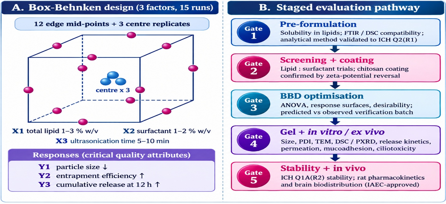
The Box–Behnken design is a three-level response-surface design in which each factor is set at low, middle and high levels and the experimental points lie at the mid-points of the edges of the design space rather than at its corners [28,29]. For three factors it needs 15 runs, made up of 12 edge mid-point combinations and three centre replicates, compared with 27 runs for a full three-level factorial, and it avoids conditions in which all factors are at their extremes at once [29]. Figure 2 (panel A) shows the geometry. A second-order polynomial model with an intercept, three linear terms, three interaction terms and three quadratic terms uses 10 coefficients, so 15 runs leave five degrees of freedom, of which two come from centre-point replicates as a measure of pure error and three test lack of fit.
Published intranasal NLC work uses exactly this layout. Kadam and colleagues optimised lurasidone NLCs with a three-factor, three-level Box–Behnken design of 15 runs (12 factorial and three centre replicates) in which solid lipid concentration, surfactant mix and sonication time were the independent variables and particle size and entrapment efficiency were the responses [20]. The optimised carrier released about 80 % of drug across goat nasal mucosa in 24 hours compared with about 50 % for the drug suspension, and after intranasal dosing in rats it gave a peak concentration of 972.78 ng/mL against 240.5 ng/mL for the plain drug, a 5.16-fold increase in bioavailability [20].
8.3Proposed factors and responses for quercetin
An analogous design for quercetin would treat total lipid concentration (for example 1 to 3 % w/v), surfactant concentration (1 to 2 % w/v) and ultrasonication time (5 to 10 minutes) as the three factors, with mean particle size (to be minimised), entrapment efficiency (to be maximised) and cumulative release at 12 hours (to be maximised) as responses. After analysis of variance, model diagnostics and desirability-based selection, a verification batch should be prepared and its measured responses compared with the predicted values. Two limits of this plan should be recognised. The design optimises the uncoated core, whereas the chitosan step adds its own variables, namely polymer molecular weight, concentration and contact time, which are best screened separately or added as factors in a second design. In addition, since the coating raises particle size [16], the size target for the core must leave room for the corona.
9Characterisation and evaluation strategy
The evaluation of the optimised formulation should move in stages, as outlined in Figure 2 (panel B) and summarised in Table 2. Particle size, polydispersity index and zeta potential are measured by dynamic and electrophoretic light scattering; transmission electron microscopy confirms shape and absence of aggregation. Entrapment efficiency and drug loading are best determined indirectly, by separating free drug by ultracentrifugation or dialysis and applying a mass balance. Differential scanning calorimetry, powder X-ray diffraction and infrared spectroscopy assess crystallinity, lipid polymorphism, drug–excipient interaction and the presence of chitosan bands on the coated surface. These analytical steps rely on a method validated to ICH Q2(R1) [15].
Release testing by dialysis or Franz diffusion cell in simulated nasal fluid gives profiles that can be fitted to zero-order, first-order, Higuchi and Korsmeyer–Peppas models to identify the release mechanism [31,32]. Ex vivo permeation across excised goat or sheep nasal mucosa in a Franz cell yields cumulative permeation, flux and an enhancement ratio against uncoated and free-drug controls, as in the lurasidone study [20]. Mucoadhesion is quantified as the force needed to detach the formulation from mucosa, again comparing coated with uncoated particles. Nasal safety is assessed by histology of mucosa exposed to the formulation, with saline as a negative control and a known ciliotoxic agent as a positive control; the coated NLCs of Gartziandia and colleagues, for instance, showed no toxicity in mouse nasal mucosa [24].
| Attribute | Method | Purpose and reference point |
|---|---|---|
| Particle size and PDI | Dynamic light scattering | Size below about 200 nm is described as suitable for the olfactory route; smaller NLCs (< 50 nm) gave higher CSF levels than > 100 nm in one study [10] |
| Zeta potential | Electrophoretic light scattering | Shift from negative to positive confirms chitosan coating (−14.4 to +20.8 mV in a Tanshinone IIA NLC) [16] |
| Morphology | TEM (SEM or AFM optional) | Shape, absence of aggregation, visible corona |
| Entrapment and loading | Indirect assay after ultracentrifugation or dialysis; HPLC or UV | Mass balance; quercetin lipid systems have reached about 78 to 96 % entrapment [3,4,22] |
| Solid-state behaviour | DSC, PXRD, FTIR | Crystallinity, polymorphism, drug–excipient compatibility, chitosan bands |
| Drug release | Dialysis or Franz cell, simulated nasal fluid; Higuchi and Korsmeyer–Peppas fitting | Release mechanism [31,32]; 83.74 % release reported for a quercetin NLC gel [4] |
| Gel properties | Gelation temperature, pH, viscosity, gel strength | Gelation about 32–34 °C [10]; pH about 4.5–6.5 [9] |
| Ex vivo permeation | Franz cell, goat or sheep nasal mucosa | Flux and enhancement ratio against uncoated NLC and free drug [20] |
| Mucoadhesion | Detachment force on excised mucosa | Coated versus uncoated particles; chitosan molecular weight effect [25] |
| Ciliotoxicity | Histology of exposed mucosa with negative and positive controls | Epithelial and ciliary integrity [24] |
| Stability | 40 ± 2 °C / 75 ± 5 % RH; size, PDI, zeta potential, content | ICH Q1A(R2) accelerated conditions [30] |
| In vivo pharmacokinetics | Wistar rats; intranasal versus intravenous; blood and brain sampling | Cmax, Tmax, AUC, DTE and DTP [4] |
Accelerated stability testing at 40 ± 2 °C and 75 ± 5 % relative humidity is specified by ICH Q1A(R2), and the guideline's accelerated study covers six months, so a shorter three-month programme should be described as preliminary [30]. In vivo work in healthy rats, approved by the institutional animal ethics committee, should compare the optimised formulation given intranasally with the free drug given intranasally and the free drug given intravenously. Brain and blood concentrations then give Cmax, Tmax and AUC, and from them the drug-targeting efficiency and direct-transport percentage, which allow results to be set against the values of 117.47 % and 88.9 % reported for a quercetin NLC gel [4].
Data from all these studies should be reported as mean ± standard deviation of at least three independent batches or animals, and compared by analysis of variance with a suitable post-hoc test or by Student's t-test where only two groups are involved, with p below 0.05 taken as significant. Because a nasal formulation is a multi-component system, each control matters: the plain (uncoated) nanoparticle isolates the effect of chitosan, the gel-free dispersion isolates the effect of the vehicle, and the free-drug solution defines the baseline that any improvement is measured against.
10Challenges and future perspectives
Several gaps limit what can be concluded from the present literature. The first is comparability. The intranasal quercetin studies in Table 1 differ in animal model, dose, endpoint and analytical method, and none has compared coated and uncoated lipid carriers of the same composition, so the specific contribution of chitosan functionalisation to nose-to-brain delivery of quercetin remains untested. Studies that include an uncoated NLC control of otherwise identical composition would answer this directly.
The second is translation. Rodent nasal anatomy differs from that of humans, and in humans the olfactory epithelium occupies only a limited part of the nasal surface [17]. Deposition therefore depends on device and droplet characteristics, and one review reports that optimal olfactory deposition is associated with particles or droplets of about 8 to 12 µm, a size range far above that of the nanoparticles themselves [9]. A formulation that performs well after pipette instillation in a rat may deposit differently from a spray in a human. Devices such as ViaNase and OptiNose are in clinical trials for brain-directed indications [10], and future quercetin work should consider the delivery device early.
The third is safety and quality. Chitosan opens tight junctions, which is useful for permeation but requires attention to long-term mucosal tolerance, and repeated dosing over weeks, as in the 14-day mouse study of Bazaz and colleagues [5], will need histological follow-up. Lipid nanoparticles can undergo polymorphic changes and size growth on storage, and the addition of a gel base and a cationic coating adds further interactions to monitor. Finally, quantification of quercetin and its conjugated metabolites in brain tissue demands a sensitive and selective analytical method, which is why the method-validation step is placed first in the evaluation pathway.
Future work should therefore integrate the elements that have so far been studied separately: a lipid core designed by statistical optimisation, a low-molecular-weight chitosan corona, a compatible in situ gel, controlled comparisons with an uncoated carrier, and pharmacokinetic and pharmacodynamic endpoints that can be compared with published values.
11Conclusion
Quercetin has antioxidant and anti-inflammatory properties relevant to neurodegeneration, but poor solubility, extensive conjugation and limited brain access hold back its use. Intranasal delivery of lipid nanoparticles has already produced encouraging results for quercetin, with entrapment of 78 to 96 % and drug-targeting efficiencies above 100 % in one study. Chitosan coating of NLCs, supported by data from other drugs, offers a way to prolong mucosal contact and to open tight junctions, and a thermosensitive gel can extend residence time further. A Box–Behnken design within a QbD framework, followed by a staged evaluation that includes mucoadhesion and ciliotoxicity, is a sound way to develop such a system. The main scientific question that remains open is whether chitosan functionalisation measurably improves nasal retention and brain delivery of quercetin compared with an unmodified lipid nanoparticle.
Conflict of interest. The authors declare that there is no conflict of interest.
Acknowledgement. The authors thank the Department of Pharmaceutics, S. N. College of Pharmacy, Jaunpur, for academic support during the preparation of this article.
Author contributions. Jaya Maurya: literature search, data compilation and drafting of the manuscript. Anoop Kumar Singh: conceptualisation, supervision and critical revision of the manuscript. Both authors have read and approved the final version.
Conflict of interest
The authors declare that there is no conflict of interest.
Acknowledgement
The authors thank the Department of Pharmaceutics, S. N. College of Pharmacy, Jaunpur, for academic support during the preparation of this article.
Author contributions
Jaya Maurya: literature search, data compilation and drafting of the manuscript. Anoop Kumar Singh: conceptualisation, supervision and critical revision of the manuscript. Both authors have read and approved the final version.
References
- [1]Middleton E Jr, Kandaswami C, Theoharides TC. The effects of plant flavonoids on mammalian cells: implications for inflammation, heart disease, and cancer. Pharmacol Rev. 2000;52(4):673–751.
- [2]Anand David AV, Arulmoli R, Parasuraman S. Overviews of biological importance of quercetin: a bioactive flavonoid. Pharmacogn Rev. 2016;10(20):84–9.
- [3]Satpathy B, Rajwar TK, Mishra A, Saha I, Rai VK, Sahoo RK, et al. Intra-nasal delivery of quercetin-loaded nanostructured lipid carriers (NLCs) in the management of Alzheimer's disease: formulation, in vitro and in vivo assessment. Mol Neurobiol. 2025;62:13182–209. https://doi.org/10.1007/s12035-025-05137-4
- [4]Sonawane D, Pokharkar V. Quercetin-loaded nanostructured lipid carrier in situ gel for brain targeting through intranasal route: formulation, in vivo pharmacokinetic and pharmacodynamic studies. AAPS PharmSciTech. 2024;25:30. https://doi.org/10.1208/s12249-024-02736-7
- [5]Bazaz MR, Chary PS, Guin S, Mehra NK, Dandekar MP. Nose-to-brain delivery of quercetin-loaded chitosan nanoparticles improves post-traumatic stress disorder-like phenotypes in mice. Eur J Pharmacol. 2026;1012:178477.
- [6]Pinheiro RGR, Pinheiro M, Neves AR. Nanotechnology innovations to enhance the therapeutic efficacy of quercetin. Nanomaterials (Basel). 2021;11(10):2658. https://doi.org/10.3390/nano11102658
- [7]Almeida AF, Borge GIA, Piskula M, Tudose A, Tudoreanu L, Valentová K, et al. Bioavailability of quercetin in humans with a focus on interindividual variation. Compr Rev Food Sci Food Saf. 2018;17(3):714–31. https://doi.org/10.1111/1541-4337.12342
- [8]Pardeshi CV, Belgamwar VS. Direct nose to brain drug delivery via integrated nerve pathways bypassing the blood-brain barrier: an excellent platform for brain targeting. Expert Opin Drug Deliv. 2013;10(7):957–72.
- [9]Drath I, Richter F, Feja M. Nose-to-brain drug delivery: from bench to bedside. Transl Neurodegener. 2025;14:23. https://doi.org/10.1186/s40035-025-00481-w
- [10]Koo J, Lim C, Oh KT. Recent advances in intranasal administration for brain-targeting delivery: a comprehensive review of lipid-based nanoparticles and stimuli-responsive gel formulations. Int J Nanomedicine. 2024;19:1767–807. https://doi.org/10.2147/IJN.S439181
- [11]Illum L. Nasal drug delivery - possibilities, problems and solutions. J Control Release. 2003;87(1–3):187–98.
- [12]Müller RH, Radtke M, Wissing SA. Nanostructured lipid matrices for improved microencapsulation of drugs. Int J Pharm. 2002;242(1–2):121–8.
- [13]Ways TMM, Lau WM, Khutoryanskiy VV. Chitosan and its derivatives for application in mucoadhesive drug delivery systems. Polymers (Basel). 2018;10(3):267. https://doi.org/10.3390/polym10030267
- [14]Pinheiro RGR, Granja A, Loureiro JA, Pereira MC, Pinheiro M, Neves AR, et al. Quercetin lipid nanoparticles functionalized with transferrin for Alzheimer's disease. Eur J Pharm Sci. 2020;148:105314.
- [15]International Council for Harmonisation. ICH Harmonised Tripartite Guideline: Validation of Analytical Procedures: Text and Methodology Q2(R1). Geneva: ICH; 2005.
- [16]Hassan DM, El-Kamel AH, Allam EA, Bakr BA, Ashour AA. Chitosan-coated nanostructured lipid carriers for effective brain delivery of Tanshinone IIA in Parkinson's disease: interplay between nuclear factor-kappa β and cathepsin B. Drug Deliv Transl Res. 2024;14(2). https://doi.org/10.1007/s13346-023-01407-7
- [17]Gänger S, Schindowski K. Tailoring formulations for intranasal nose-to-brain delivery: a review on architecture, physico-chemical characteristics and mucociliary clearance of the nasal olfactory mucosa. Pharmaceutics. 2018;10(3):116. https://doi.org/10.3390/pharmaceutics10030116
- [18]Naseri N, Valizadeh H, Zakeri-Milani P. Solid lipid nanoparticles and nanostructured lipid carriers: structure, preparation and application. Adv Pharm Bull. 2015;5(3):305–13.
- [19]Dhawan S, Kapil R, Singh B. Formulation development and systematic optimization of solid lipid nanoparticles of quercetin for improved brain delivery. J Pharm Pharmacol. 2011;63(3):342–51. https://doi.org/10.1111/j.2042-7158.2010.01225.x
- [20]Kadam T, Agrawal S, Shetty S. Novel nanostructured lipid carriers with lurasidone hydrochloride for intranasal administration for improved bioavailability. Ther Deliv. 2025. https://doi.org/10.1080/20415990.2025.2477440
- [21]Revankar AA, Patil AS, Karishetti R, Chougule KR, Patil P, Salokhe A. Enhanced bioavailability of quercetin-loaded niosomal in situ gel for the management of Parkinson's disease. Front Pharmacol. 2025;15:1519649. https://doi.org/10.3389/fphar.2024.1519649
- [22]Elkomy MH, Zaki RM, Alsaidan OA, Elmowafy M, Zafar A, Shalaby K, et al. Intranasal nanotransferosomal gel for quercetin brain targeting: I. Optimization, characterization, brain localization, and cytotoxic studies. Pharmaceutics. 2023;15(7):1805. https://doi.org/10.3390/pharmaceutics15071805
- [23]Popescu R, Ghica MV, Dinu-Pîrvu CE, Anuța V, Lupuliasa D, Popa L. New opportunity to formulate intranasal vaccines and drug delivery systems based on chitosan. Int J Mol Sci. 2020;21(14):5016. https://doi.org/10.3390/ijms21145016
- [24]Gartziandia O, Herran E, Pedraz JL, Carro E, Igartua M, Hernandez RM. Chitosan coated nanostructured lipid carriers for brain delivery of proteins by intranasal administration. Colloids Surf B Biointerfaces. 2015;134:304–13.
- [25]Bruinsmann FA, Pigana S, Aguirre T, Dadalt Souto G, Pereira GG, Bianchera A, et al. Chitosan-coated nanoparticles: effect of chitosan molecular weight on nasal transmucosal delivery. Pharmaceutics. 2019;11(2):86. https://doi.org/10.3390/pharmaceutics11020086
- [26]International Council for Harmonisation. ICH Harmonised Tripartite Guideline: Pharmaceutical Development Q8(R2). Geneva: ICH; 2009.
- [27]Yu LX. Pharmaceutical quality by design: product and process development, understanding, and control. Pharm Res. 2008;25(4):781–91.
- [28]Box GEP, Behnken DW. Some new three level designs for the study of quantitative variables. Technometrics. 1960;2(4):455–75.
- [29]Ferreira SLC, Bruns RE, Ferreira HS, Matos GD, David JM, Brandão GC, et al. Box-Behnken design: an alternative for the optimization of analytical methods. Anal Chim Acta. 2007;597(2):179–93.
- [30]International Council for Harmonisation. ICH Harmonised Tripartite Guideline: Stability Testing of New Drug Substances and Products Q1A(R2). Geneva: ICH; 2003.
- [31]Higuchi T. Mechanism of sustained-action medication. Theoretical analysis of rate of release of solid drugs dispersed in solid matrices. J Pharm Sci. 1963;52(12):1145–9.
- [32]Korsmeyer RW, Gurny R, Doelker E, Buri P, Peppas NA. Mechanisms of solute release from porous hydrophilic polymers. Int J Pharm. 1983;15(1):25–35.